Microsoft Word - Text.docx
|
|
|
- えりか あさま
- 7 years ago
- Views:
Transcription
1 論 種別 : 症例報告 タイトル : 機械的 栓回収療法後に出 発症した後天性 pial AV fistula の 例 1) 1) 著者 : 進藤孝 郎荻野達也 1) 1) 1) 憲司 俊明中村博彦 1) 中村記念病院脳神経外科 渕崎智紀 1) 遠藤英樹 1) 丸賀庸平 1) 渡部寿 1) 上 連絡著者 : 進藤孝 郎 ( しんどうこういちろう ) 所属 : 医仁会中村記念病院脳神経外科住所 : 北海道札幌市中央区南 1 条 14 丁 291 番地 TEL: / FAX: [email protected] Key Words: pial arteriovenous fistula, adult, mechanical thrombectomy 本論 を, 本脳神経 管内治療学会機関誌 Journal of Neuroendovascular Therapy に投稿するにあたり, 筆頭著者, 共著者によって, 国内外の他雑誌に掲載ないし投稿されていないことを誓約致します.
2 Abstract: 中 脳動脈閉塞に対し 栓回収療法を施 し 関連する 管領域に発 した Pial arteriovenous fistula (Pial AVF) を経験したので報告する 症例 :72 歳男性 201X 年 右脳梗塞 右 M1 閉塞に対して Penumbra system による 栓回収療法を施 し mtici 2b( M2 anterior trunk は閉塞のまま ) の再開通を得た 1 週間後の MRA 検査では完全再開通しており 経過は良好で 宅退院となったが 201X+5 年に痙攣を起こし当院へ救急搬送された MRI 検査で右中 前回に 質下出 を認め DSA 検査を ったところ Rt. central artery と Vein of Trolard の間に以前は認めなかった AV shunt が確認された nidus を認めず pial AVF と診断し 開頭術による shunt point の遮断を った 術中所 から 栓回収療法後に じた 質静脈の 栓化が発症の契機となったものと推察された < 諸 > Pial arteriovenous fistula (Pial AVF) は 較的稀な 管病変である 以前は arteriovenous malformation(avm) の subtype に分類されていた 1) が 動静脈間に nidus を伴わず徐々に独 して認識されるようになった 病態は未だ 分には明らかとなっていないが 成 例は出 発症が多く 2) 出 発症例の保存的加療は予後不良との報告もあり 3) 積極的な治療が必要と考えられている 今回われわれは 右中 脳動脈閉塞に対し Penumbra システム (Penumbra Inc. Alameda, CA, USA) による機械的 栓回収療法を施 し 関連する 管領域に後天性に発 した pial AVF を経験したので 献的考察
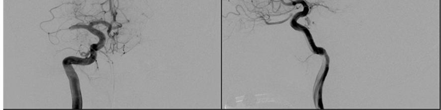
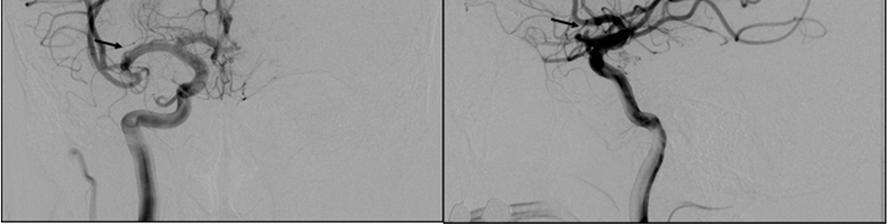
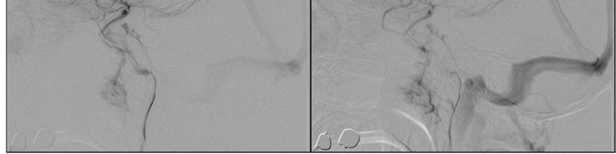
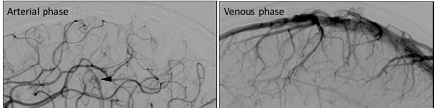
3 を含め報告する < 症例呈 > 症例 :72 歳 男性 既往歴 : 発作性 房細動 脳梗塞現病歴 :201X 年 Y 右脳梗塞 右中 脳動脈閉塞 (M1 閉塞 ) に対して rt-pa 静注療法および Penumbra システムによる 栓回収療法を施 した ( Figure 1a) Aspiration pomp による吸引をかけ 適宜 Separator を いながら Penumbra 4MAX reperfusion catheter を閉塞部位まで先進させ 栓を回収し TICI 2b( M2 anterior trunk は閉塞のまま ) の再開通を得た (Figure 1b) 再開通時の Rt. ICAGでは Vein of Trolard は Superior sagittal sinus(sss) から Superficial middle cerebral vein(smcv) 向へ下 する 還流であった (Figure 1c) 栓回収療法の 1 週間後の MRA 検査では M2 anterior trunk の再開通も確認され 施術後の経過は良好で 1 ヶ 間の 院 加療を経て modified Rankin scale (mrs) 1 で 宅退院となった この時点では心房細動が検出されておらず 二次予防薬としてはワルファリンが選択された 外来通院にて経過をみていたが 201X+5 年 Z に痙攣を起こし当院へ救急搬送された 頭部 MRI CT 検査で右前頭葉 ( 中 前回 ) 質下出 を認め (Figure 2a) 院となった DSA 検査を ったところ Rt. central artery と Vein of Trolard の間に 201X 年の 栓回収施 時には認めなかった Arteriovenous shunt(av shunt) が疑われた 出 部が中 前回であったが症状は軽度であった 2 ヶ 後に った DSA 検査で より AV shunt が明瞭化しており 動静脈間に nidus を認めなかった事から pial AVF と診
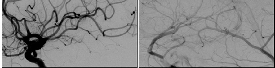
4 断 (Figure 2b) し 再出 予防のため開頭術による shunt point の遮断を計画した 術前はワルファリンを休薬し ヘパリン置換を った 術所 (Figure 3a):shunt point を中 に craniotomy を った 硬膜を切開すると AV shunt が存在する部位のくも膜は肥厚し 濁していた ヘモジデリンの沈着もあり 陳旧性のくも膜下出 の存在を 唆する所 を認めた AV shunt 周囲のくも膜を剥離し 管構造の denude を った Vein of Trolard には 眼的に正常灌流を経た静脈 と AV shunt を経由した動脈 が混在していた AV shunt は Precentral vein(vein of Trolard) に合流する cortical vein に存在しており 罹患静脈は venous pouch を形成していた Feeding artery となっている Central artery と Precentral artery から細い動脈が venous pouch に流 していた 罹患静脈は central sulcus の深部まで続いており nidus は認めなかった 術前の診断通り pial AVF と考えられた AV shunt は罹患 質静脈ごと周囲組織から離断した 術中所見として罹患 質静脈深部に静脈の 栓化と考えられる所 がみられた (Figure3b) 罹患 質静脈を剥離し摘出する過程で 陳旧性 質下出 の 腫腔内に到達し pial AVF が脳出 の原因として 盾がないものと考えられた ICG 蛍光 管造影検査および術中脳 管造影検査にて AV shunt の消失を確認し 術を終了した 術後の DSA 検査 :AV shunt は消失しており AV shunt のあった部位を境に Precentral vein は上 性 (SSS 向 ) に還流 向が変化しており SMCV は下 性のままであった (Figure 4) 術前に発作性 房細動が 電図で検出されたことから 出 性合併
5 症がないことを確認した後 DOAC(Direct oral anticoagulant) に変 更した 術後経過は良好で mrs 1 で退院となった < 考察 > Pial AVF は 1 本もしくは複数の pial artery を feeder として 通常 1 本の cortical vein を drainer としており nidus を有する AVM とは異なる頭蓋内動静脈短絡疾患である その頻度は頭蓋内動静脈短絡疾患の 1.6~4.7% 4)5) と 較的稀な疾患である Pial AVF は先天性のものと後天性のものがあり 先天性のものは HHT や CM-AVM とい った遺伝性疾患との関連 6)7) が 児例で報告があり 後天性のもの 8) 9) は頭部外傷 術あるいは静脈洞 栓症後に 過性の静脈圧亢進が起こる事で じると考察されている 発症の契機としては mass effect や出 痙攣 不全などがあり 痙攣発症は 15 歳以下の 児例で多く 出 発症は 16 歳以上の成 例で多いと Yang WH らにより報告されている 2) 静脈洞 栓症を伴う 出 性の pial AVF で抗凝固療法のみで改善 を認めたとの報告もある 10) が 出 例においては保存的治療では 63% が死亡しているとの報告がある 11) Yang WH らは成 発症例は動脈圧に対し緩衝作 のある varix を有さない例が多く 出 発症が多いと考察しており 2) 成 発症例や出 発症例においては早期に根治的な治療が必要と考えられる AVM と異なり nidus が存在しないため 病変部の除去は必ずしも必要ではなく shunt point の遮断のみで 分とされている 2) Pial AVF の治療には 直達術や 管内治療または両者の併 がある 表在性
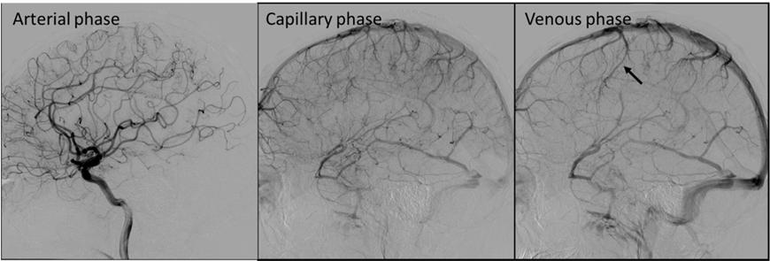
6 の病変に対しては いずれの治療法でも可能と考えられるが 深部の病変や eloquent area に存在するなど直達が困難な病変に関しては 管内治療が有利と考えられる 管内治療に いられた塞栓 12) 物質としては balloon や coil glue poly alcohol Onyx の報告 ~15) があるが shunt point が完全に遮断できないまま draining vein に塞栓物質が流 し venous out flow の停滞を招いた場合は遅発性の出 などのリスクがあるものと考えられる また 脳外の遠位塞栓にも留意が必要である Yang ら 2) の review によると直逹術による obliteration rate が 96.8% に対して 管内治療が 86.5% と根治性に関しては直達術の が優位と考えられるが 治療法選択には病変の局在や shunt point への approach の難易度など詳細な術前検討に基づいて決定する必要がある Matsubara らは摘出した 栓化 AVM の病理検査で nidus 内に 細 管を じていた事を報告し 静脈 栓を じると再開通 再灌流のため angiogenesis が誘導され 動静脈短絡の形成に関与していると考察されている 16) これまで静脈洞 栓症に伴う pial AVF 発症の報告はあるが 機械的 栓回収療法後の pial AVF 発症は渉猟し得た限りでは認めなかった Matsubara らは Superior sagittal sinus thrombosis の follow up 中に dural AVF pial AVF を じた症例を報告している 16) が 閉塞した cortical vein が再開通した部分に pial AVF が じていた 本症例では 栓回収療法の1 週間後の MRA 検査で M2 anterior trunk の再開 通が確認された 動脈の再開通までの数 の間に cortical vein
7 (draining vein) に閉塞が じたものと推察された 閉塞した cortical vein に対する 体反応として angiogenesis が誘導され そこに動脈の再開通が起こることにより閉塞した cortical vein に局所的な圧上昇が加わり pial AVF を形成した可能性が考えられた (Figure 5) 急性期再開通療法が増加している昨今において 稀ながら pial AVF は閉塞 管 配領域に起こりうる合併症として留意すべきと考えられた MRI MRA 検査等の定期検査での微細な変化にも注視する必要がある < 結語 > 機械的 栓回収療法から5 年後に出 発症した pial AVF を報告した 栓回収療法後に じた静脈灌流の変化に伴う局所的な静脈圧の亢進が発症の契機となった可能性が 唆された < 利益相反開 > 筆頭著者および共著者全員が利益相反はない References: 1)Yasargil M. Microneurosurgery ⅢA: AVM of the brain, history, embryology, pathological considerations, hemodynamics, diagnostic studies, microsurgical anatomy. Stuttgart: Georg Thieme Verlag, ) Yang WH, Lu MS, Cheng YK, et al. Pial arteriovenous fistula: a review of literature. Br J Neurosurg 2011; 25:
8 3)Nelson PK, Niimi Y, Lasjaunias P, et al: Endovascular embolization of congenital intracranial pial arterio- venous fistulas. Neuroimaging Clin N Am 1992; 2: ) Lv X, Li Y, Jiang C, Wu Z: Endovascular treatment of brain arteriovenous fistulas. AJNR Am J Neuroradiol 2009;30: ) Lv X, Jiang C, Li Y, et al. Clinical Outcomes of Endovascular Treatment for Intracranial Pial Arteriovenous Fistulas. World Neurosurg 2010; 73: ) Kikuchi K, Kowada M, Sasajima H: Vascular malformations of the brain in hereditary hemorrhagic telangiectasia (Rendu-Osler-Weber disease). Surg Neurol 1994;41: ) Ishiguro T, Komiyama M, Terada A, et al. Pial arteriovenous fistulas. Niche Neuro-Angiology Conference )Nomura S, Ishikawa O, Tanaka K, et al.pial arteriovenous fistula caused by trauma: A case report. Neurol Med Chir(Tokyo)2015:55, )Acquired pial and dural arteriovenous fistulae following superior sagittal sinus thrombosis in patients with protein S deficiency: A report of two cases. Neurol Med Chir(Tokyo) 2014;54, ) 美帆, 輪島 介, 他 : 抗凝固療法により改善を認めた静脈洞 栓症を伴う Pial AV fistulae の 1 例 : 脳神経外科速報 2016;vol.26 no ) Nelson PK, Niimi Y, Lasjaunias P, et al.endovascular embolization of congenital intracranial pial arterio- venous fistulas.
9 Neuroimaging Clin N Am 1992;2: ) Vinuela F, Drake CG, Fox AJ, Pelz DM. Giant intracranial varices secondary to high-flow arteriovenous fistulae. J Neurosurg. 1987; 66: ) Vinuela F, Fox AJ, Kan S, Drake CG. Balloon occlusion of a spontaneous fistula of the posterior inferior cerebellar artery. Case report. J Neurosurg. 1983; 58: ) Wang YC, Wong HF, Yeh YS. Intracranial pial arteriovenous fistulas with single-vein drainage. Report of three cases and review of the literature. J Neurosurg Feb; 100(2 suppl Pediatrics): ) Kim HM, Cho JH, Kim KH. Onyx embolization of intracranial pial arteriovenous fistula. J cerebrovasc endovasc neurosurg. 2016; 18: ) Matsubara S, Satoh K, Satomi J, et al: Acquired pial and dural arteriovenous fistula following superior sagittal sinus thrombosis in patients with protein S deficiency: a report of two cases. Neurol Med Chir (Tokyo) 2014; 54:
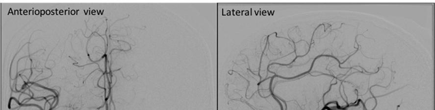
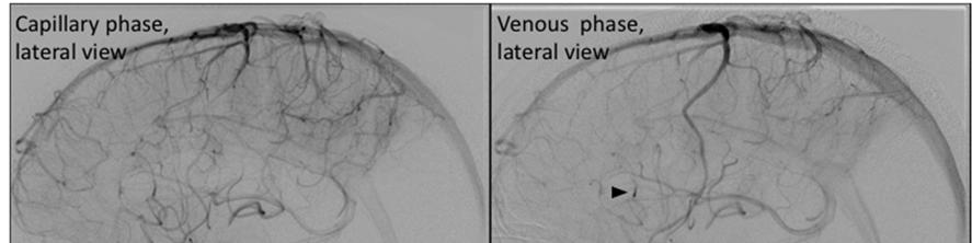
10 Figure Legends: Figure 1a. Right internal carotid angiography showed occlusion at the M1 segment of the middle cerebral artery. Figure 1b. Penumbra reperfusion catheter was advanced to the occlusion site and thrombus was removed. Right internal carotid angiography showed the right M2 anterior trunk occlusion (arrow) after multiple pass. (TICI 2b) Figure 1c. Right internal carotid angiography of post thrombectomy. Due to a residual thrombus, contrast medium stagnated at the right M2 anterior trunk (Arrowhead). The flow of precentral vein was descending from SSS to SMCV. Figure 2a. Axial plain CT showed subcortical hemorrhage in the right precentral gyrus. Figure 2b. Right internal carotid angiography showed a pial arteriovenous fistula with feeding artery arising from central artery. A pial arteriovenous fistula drained to the vein of Trolard (Arrowhead).
11 Figure 3a. Intraoperative photograph showed precentral vein (the vein of Trolard) changed red vein(arrows) and there was a venous pauch in the side of precentral gyrus. Feeding arteries assembled to the venous pouch. (arrowhead) Figure 3b. Intraoperative photograph. The vessel (arrow) in deep portion of central sulcus was recognized as thrombosed vein. Figure 4. Post-operative right carotid angiogram showing complete obliteration of the fistula. The flow of precentral vein was ascending to SSS. (arrow) Figure 5. Schematic drawing explaining a possible mechanism for pial AVF development. A: Normal venous perfusion. The cortical veins from frontal lobe perfused to SSS and those from parietal lobe to SMCV. B: During of M2 anterior trunk occlusion, venous perfusion pressure from frontal lobe was lower and the cortical vein seemed to be thrombosed.
12 C: After recanalization of M2 anterior trunk, venous perfusion pressure from frontal lobe was recovered and venous pressure of the occluded vein locally increased. D: When the patient bled in the precentral gyrus, the pial AVF was indicated with drainage to the cortical vein connecting to the vein of Trolard.

13 Figure 1a. Figure 1b.
14 Figure 1c.

15 Figure 2a. Figure 2b.
 Intraoperative view 2) Color")

16 Figure 3a. 1) Intraoperative view 2) Color map during ICG injection 3) Intraoperative view after removal arachnoid membrane Figure 3b.
17 Figure 4.
18 Figure 5.
1 Case report 後硬膜動脈から延髄外側への穿通枝を認めた 1 例 A case of Lateral medullary artery arise from the Posterior meningeal artery 伊藤真史 泉孝嗣 西堀正洋 今井資 玉利洋介 塚田哲也 石田衛 クロ
 1 Case report 後硬膜動脈から延髄外側への穿通枝を認めた 1 例 A case of Lateral medullary artery arise from the Posterior meningeal artery 伊藤真史 泉孝嗣 西堀正洋 今井資 玉利洋介 塚田哲也 石田衛 クロップ明日香 若 林俊彦 Masashi I, Takashi I, Nishihori M, Tasuku
6) キーワード 頭蓋頸椎移行部 硬膜動静脈瘻 クモ膜下出血 血管内治療 開頭手術 7) 宣言 本論文を 日本脳神経血管内治療学会 機関誌 JNET journal of Neuroendvascular Therapy に投稿するにあたり 筆頭著者 共著者に よって 国内外の他雑誌に掲載ないし投稿
 キーワード 頭蓋頸椎移行部 硬膜動静脈瘻 クモ膜下出血 血管内治療 開頭手術 7) 宣言 本論文を 日本脳神経血管内治療学会 機関誌 JNET journal of Neuroendvascular Therapy に投稿するにあたり 筆頭著者 共著者に よって 国内外の他雑誌に掲載ないし投稿") 1) 論文種別症例報告 2) 論文タイトル 血管内治療を先行し開頭術を追加して治療した 頭蓋頸椎移行部硬膜動 静脈瘻の一例 3) 全員の著者名 金子純也 1 柴田あみ 1 北橋章子 1 工藤小織 1 畝本恭子 1 山口英宣 2 山崎道生 3 玉置智規 3 松本学 4 横田裕行 5 6 兵頭明夫 4) 1 日本医科大学多摩永山病院救命救急センター 2 同放射線科 3 同脳神経外科 4 山梨県立中央病院救命救急センター
1) 論文種別症例報告 2) 論文タイトル 血管内治療を先行し開頭術を追加して治療した 頭蓋頸椎移行部硬膜動 静脈瘻の一例 3) 全員の著者名 金子純也 1 柴田あみ 1 北橋章子 1 工藤小織 1 畝本恭子 1 山口英宣 2 山崎道生 3 玉置智規 3 松本学 4 横田裕行 5 6 兵頭明夫 4) 1 日本医科大学多摩永山病院救命救急センター 2 同放射線科 3 同脳神経外科 4 山梨県立中央病院救命救急センター
Kittipong
 1. Aoki R, Srivatanakul K, Hirayama A, A vein of the foramen caecum observed on angiography, Eur J Ana 21(4): 305-307, 2017 2. Hui Han & Wei Tao & Ming Zhang, The dural entrance of cerebral bridging veins
症例_佐藤先生.indd
 症例報告 JNET 7:259-265, 2013 後拡張手技を行わない頚動脈ステント留置術後の過灌流状態においてくも膜下出血とステント閉塞を来した 1 例 Case of Subarachnoid Hemorrhage and In-Stent Occlusion Following Carotid rtery Stenting without Post alloon Dilatation ccompanied
Table 1 ICHs in young adults Table 2 Five cases of undetermined etiology
 Table 1 ICHs in young adults Table 2 Five cases of undetermined etiology 神 1 1. left : in the right ted MRI, callosum. parietal. right : ICH in 保 洋 之 他 ICHs 4 Ti-weighthe 1. left: rrhagic corpus infarction
石黒
 Fig.3: Transcerebral route 3A: 上衣下静脈は transcerebral veinを介して 脳表の静脈と交通する 3B C: 脳深部静脈血栓症の症例 両側ICV BVRとガレン大静脈 直静脈洞は閉塞している 左内頚動脈 撮影 静脈相の正面像 (3B)と側面像 (3C) 深部静脈の血流は側脳室体部や三角部の外側壁から3本の transcerebral veinを介してvein
脈管学55巻11号 pp
 Online publication December 10, 2015 191 原 著 55 21 1 2 2 1 2 2 2 2 1 要旨 : 8 21 19 2 4 1 20 SMA short segment J Jpn Coll Angiol 2015; 55: 191 196 Key words: superior mesenteric artery, dissection, conservative
Spinal Arteriovenous Shunts Which are Curable and which Incurable? Yuji Matsumaru, M.D., PhD. 1, Takayuki Hara, M.D. 2, and Akira Matsumur
 1 2 3 1 2 3 Spinal Arteriovenous Shunts Which are Curable and which Incurable? Yuji Matsumaru, M.D., PhD. 1, Takayuki Hara, M.D. 2, and Akira Matsumura, M.D. 3 1 Department of Endovascular Neurosurgery,
全国循環器撮影研究会 HP講座No
 HP No.6 4 HP No.6 4 NTT 1. 1936 1953 Seldinger Seldinger 1956 Odman X Interventional Radiology IVR 1967 Margulis 1964 Dotter 2. 2.1 IVR CT MR IVR 2.2-1 - HP No.6 4 2.3 feeding artery parasitic supply increased
JNET 9: , 2015 症例報告 スーパー政宗 ( 第二報 ) - 初期臨床経験 - Super-Masamune: initial clinical experience Masayuki EZURA, Naoto KIMURA, Hiroshi UENOHARA Departm
 - 初期臨床経験 - Super-Masamune: initial clinical experience Masayuki EZURA, Naoto KIMURA, Hiroshi UENOHARA Departm") JNET 9:192 196, 2015 症例報告 スーパー政宗 ( 第二報 ) - 初期臨床経験 - Super-Masamune: initial clinical experience Masayuki EZURA, Naoto KIMURA, Hiroshi UENOHARA Department of Neurosurgery, NHO Sendai Medical Center Abstract
JNET 9:192 196, 2015 症例報告 スーパー政宗 ( 第二報 ) - 初期臨床経験 - Super-Masamune: initial clinical experience Masayuki EZURA, Naoto KIMURA, Hiroshi UENOHARA Department of Neurosurgery, NHO Sendai Medical Center Abstract
Microsoft PowerPoint - Horie review
 急性期脳梗塞治療における血管内治療の重要性 ~Trevo R XP ProVue Retriever の実力 ~ 急性期脳梗塞治療に対するチーム連携と挑戦 長崎大学脳神経外科堀江信貴 筆頭演者は日本脳神経外科学会への COI 自己申告を完了しております 本演題の発表において開示すべき COI はありません Jeffrey L. Saver et al. Stroke.2006;37:263-266.
HO_edit 脳梗塞の血管内治療.pptx
 脳梗塞の血管内治療 N Engl J Med 2015; 372:11-20 N Engl J Med 2015; 372:1019-1030 N Engl J Med 2015; 372:1009-1018 慈恵 ICU 勉強会 2015 年 3 月 31 日 石垣昌志 急性期脳梗塞治療戦略の基本概念 閉塞している血管の再開通 ペナンブラ領域を救済 予後が改善 慈恵 ICU 火曜勉強会脳梗塞ガイドライン
Microsoft Word - Text.docx
 1) 論文種別テクニカルノート 2) 論文タイトル上矢状静脈洞血栓症に対し coaxial catheter からの血栓吸引が有効であった一例 3) 著者名藤井照子 1) 芳村雅隆 2) 廣田晋 2) 清川樹里 3) 山本信二 2) 4) 著者の所属施設 部署 1) 青梅市立総合病院脳神経外科 2) 土浦協同病院脳神経外科 3) マサチューセッツ総合病院脳神経外科 5) 連絡著者の氏名 連絡先 氏名芳村
FLONTA Vol.2 FlowGate 2 Balloon Guide Catheter technical assistant FlowGate 2 Balloon Guide Catheter を使用した臨床経験 佐世保市総合医療センター脳神経外科 林健太郎先生 FlowGate 2 Bal
 FLONT Vol.2 FlowGate 2 alloon Guide Catheter technical assistant FlowGate 2 alloon Guide Catheter 佐世保市総合医療センター脳神経外科 林健太郎先生 FlowGate 2 alloon Guide Catheter 8Fr 0.084inches 5 FlowGate 2 P002164.v.1.0 Page
日本職業・災害医学会会誌第51巻第5号
 太田ら 同時多発性高血圧性脳葉出血の 1 手術例 図 1 搬入時 CT 上段 単純 CT 下段 造影 CT 図 2 術後の脳血管撮影検査では明らかな出血源は認めない 379 Reprint request: MULTIPLE SIMULTANEOUS HYPERTENSIVE LOBAR HEMORRHAGE Hirotsugu OHTA M.D. 1) and Akira YOKOTA M.D.
論 種別 :Case report Title: バルーンカテーテルを活 し TAE TVE で根治した横静脈洞の静脈洞交会近傍部からS 状静脈洞硬膜動静脈瘻の1 例 著者名 Hajime Yabuzaki 1, Tomoaki Terada 2, Hisato Ikeda 3, Michiari
 論 種別 :Case report Title: バルーンカテーテルを活 し TAE TVE で根治した横静脈洞の静脈洞交会近傍部からS 状静脈洞硬膜動静脈瘻の1 例 著者名 Hajime Yabuzaki 1, Tomoaki Terada 2, Hisato Ikeda 3, Michiari Kawamo 3, Akira Wada 1, Sadayoshi Nakayama 1, Tomomi
本論文を 日本脳神経血管内治療学会機関誌 JNET Journal of Neuroendovascular Therapy に投稿するにあたり, 筆頭著者, 共著者によっ て, 国内外の他雑誌に掲載ないし投稿されていないことを誓約致します.
 論文種別 : 症例報告 論文タイトル : 顔面静脈経由に Distal Access Catheter を海綿静脈洞内へ留置し 治療した 海綿静脈洞部硬膜動静脈瘻の 1 例 著者名 : 千原英夫波多野武人定政信猛甲斐康稔坂真人安藤充重瀧田亘 徳永敬介鎌田貴彦永田泉 所属施設 部署 : 平成紫川会小倉記念病院脳神経外科 連絡著者の氏名 連絡先 : 氏名 : 千原英夫 所属 : 平成紫川会小倉記念病院脳神経外科
et al No To Shinkei Clin Neurol Neurology et al J Neurosurg et al Arch Neurol et al Angiology
 37 Recommendations for the Management of Moyamoya Disease A Statement from Research Committee on Spontaneous Occlusion of the Circle of Willis (Moyamoya Disease) Research on intractable diseases of the
症例_原先生.indd
 症例報告 JNET 7:266-274, 2013 破裂脳底動脈分岐部動脈瘤に対して Y ステントテクニックを用いて瘤内コイル塞栓術を施行した 2 例 1 1 1 1 1 2 2 2 Y-configured Stent-assisted Coil Embolization of Basilar Bifurcation Aneurysms: report of 2 cases Takeshi HARA
GE ヘルスケア ジャパン 3D ASL( 非造影頭部灌流画像 ) の実践活用 IMS( イムス ) グループ医療法人社団明芳会横浜新都市脳神経外科病院画像診療部竹田幸太郎 当院のご紹介横浜新都市脳神経外科病院 ( 横浜市 青葉区 ) は 1985 年に開院し 患者さんの 満足 と 安心 を第一に考
 の実践活用 IMS( イムス ) グループ医療法人社団明芳会横浜新都市脳神経外科病院画像診療部竹田幸太郎 当院のご紹介横浜新都市脳神経外科病院 ( 横浜市 青葉区 ) は 1985 年に開院し 患者さんの 満足 と 安心 を第一に考") GE ヘルスケア ジャパン 3D ASL( 非造影頭部灌流画像 ) の実践活用 IMS( イムス ) グループ医療法人社団明芳会横浜新都市脳神経外科病院画像診療部竹田幸太郎 当院のご紹介横浜新都市脳神経外科病院 ( 横浜市 青葉区 ) は 1985 年に開院し 患者さんの 満足 と 安心 を第一に考え 愛し愛される病院を目指す を病院理念に基づき 地域医療の貢献に努力し続けている クモ膜下出血 脳出血
GE ヘルスケア ジャパン 3D ASL( 非造影頭部灌流画像 ) の実践活用 IMS( イムス ) グループ医療法人社団明芳会横浜新都市脳神経外科病院画像診療部竹田幸太郎 当院のご紹介横浜新都市脳神経外科病院 ( 横浜市 青葉区 ) は 1985 年に開院し 患者さんの 満足 と 安心 を第一に考え 愛し愛される病院を目指す を病院理念に基づき 地域医療の貢献に努力し続けている クモ膜下出血 脳出血
テクニカルノート JNET 2: , 2008 海綿静脈洞部硬膜動静脈瘻の血管内治療において 3D-Rotational Angiography が有用であった 1 例 良好な 3D 画像を得るための工夫 東 1) 登志夫 1) 中原一郎 1,3) 松本省二 1) 岩室康司 1) 渡邉芳
 登志夫 1) 中原一郎 1,3) 松本省二 1) 岩室康司 1) 渡邉芳") テクニカルノート JNET 2:232-237, 2008 海綿静脈洞部硬膜動静脈瘻の血管内治療において 3D-Rotational Angiography が有用であった 1 例 良好な 3D 画像を得るための工夫 東 登志夫 中原一郎 1,3) 松本省二 岩室康司 渡邉芳彦 武澤正浩 村田大樹 1,4) 岩朝光利 1,5) 森谷淳二 2) 一ノ瀬良二 A case of successful endovascular
テクニカルノート JNET 2:232-237, 2008 海綿静脈洞部硬膜動静脈瘻の血管内治療において 3D-Rotational Angiography が有用であった 1 例 良好な 3D 画像を得るための工夫 東 登志夫 中原一郎 1,3) 松本省二 岩室康司 渡邉芳彦 武澤正浩 村田大樹 1,4) 岩朝光利 1,5) 森谷淳二 2) 一ノ瀬良二 A case of successful endovascular
症例_一ノ瀬先生.indd
 症例報告 JNET 7:317-322, 2013 右側大動脈弓を有する破裂脳動脈瘤症例に対して脳血管内治療を行った 1 例 1 2 1 1 1 2 2 1 1 case of ruptured cerebral aneurysm with a right-sided aortic arch treated by coil embolization Nobuhiko ICHINOSE 1,2) Yoshihiro
症例_森谷先生.indd
 症例報告 JNET 5:195-201, 2012 FilterWire EZ を用いたステント留置術中に no flow を来した 1 例 : 症例報告 No flow phenomenon during carotid artery stenting with the use of FilterWire EZ: a case report Masao MORIY Hiroshi ITOKW Michio
Part 1 症状が強すぎて所見が取れないめまいをどうするか? 頭部 CT は中枢性めまいの検査に役立つか? 1 めまい診療が難しい理由は? MRI 感度は 50% 未満, さらには診断学が使えないから 3
 Part 1 画像に頼らない, 明日から使えるめまい診察伝授 1 めまい診療が難しい理由は? MRI 感度は 50% 未満, さらには診断学が使えないから 症例 1 症例 1 めまい 50 歳女性 起床時からめまいがあり, 改善しないため救急要請 搬送後にストレッチャーへ移動したとたん嘔吐 症状が続き非常に辛そうで, 問診はほとんどできない 身体所見を取ることも難しい バイタルサインは安定している
MCIS Vol.2 The Most Conformable Intracranial Stent 財団法人平成紫川会小倉記念病院脳神経外科 太田剛史先生 Neuroform EZ Stent System open cell closed cell Neuroform open cell Jac
 The Most Conformable Intracranial Stent 財団法人平成紫川会小倉記念病院脳神経外科 太田剛史先生 open cell closed cell Neuroform open cell Jack Up Neuroform EZ Stent System Neuroform EZ Neuroform EZ STENT SYSTEM ible Design. Enhanced
くろすはーと30 tei
 1No.30 017 1 脳神経内科 脳神経内科部長 北山 次郎 脳神経外科部長 吉岡 努 皆様へお知らせです 既にお気づきの方もおられる 高脂血症など生活習慣病を背景とした脳血管病変の 013年4月に脳血管内手術を当院に導入するために 代表的な手術として 脳動脈瘤の手術 動脈瘤コイル塞 かとは思いますが このたび016年10月より当院脳 評価や治療にあたる一方で 意識障害 けいれん 頭 赴任し 脳血管内手術の定着のために業務上の調整を
レイアウト 1
 北海道脳神経疾患研究所医誌第 24 巻 2013.12.P23 27 当院における Door to Puncture time(d2p) 短縮についての考察 高平一樹 片岡丈人 荻野達也 遠藤英樹 中村博彦中村記念病院脳神経外科脳血管内治療センター Considerations to reduce the time from door to puncture (D2P) in our hospital
テクニカル_田中先生.indd
 テクニカルノート JNET 5:202-207, 2012 遺残三叉神経動脈分岐部に生じた大径内頚動脈瘤に対する tandem balloon による balloon test occlusion 1 1 1 2 1 1 1 3 1 1 1 Tandem balloon test occlusion for a large unruptured aneurysm associated with persistent
症例報告 JNET 2: , 2008 Anterior condylar confluent 近傍硬膜動静脈瘻の 2 例 1) 佐々木哲郎 2) 長島久 2) 佐藤大輔 2) 小山淳一 1) 本郷一博 Dural arteriovenous fistula around the ant
 佐々木哲郎 2) 長島久 2) 佐藤大輔 2) 小山淳一 1) 本郷一博 Dural arteriovenous fistula around the ant") 症例報告 JNET 2:212-216, 2008 Anterior condylar confluent 近傍硬膜動静脈瘻の 2 例 1) 佐々木哲郎 長島久 佐藤大輔 小山淳一 1) 本郷一博 Dural arteriovenous fistula around the anterior condylar confluent: Report of two cases Tetsuo SASAKI
症例報告 JNET 2:212-216, 2008 Anterior condylar confluent 近傍硬膜動静脈瘻の 2 例 1) 佐々木哲郎 長島久 佐藤大輔 小山淳一 1) 本郷一博 Dural arteriovenous fistula around the anterior condylar confluent: Report of two cases Tetsuo SASAKI
脳卒中の医療連携体制を担う医療機関等における実績調査 調査内容 平成 28 年度の実績 ( 調査内容は別紙様式のとおり ) 別紙 1: 急性期の医療機能を有する医療機関用別紙 2: 急性期及び回復期の医療機能を有する医療機関用別紙 3: 回復期の医療機能を有する医療機関用別紙 4: 維持期の医療機能
 別紙 1: 急性期の医療機能を有する医療機関用別紙 2: 急性期及び回復期の医療機能を有する医療機関用別紙 3: 回復期の医療機能を有する医療機関用別紙 4: 維持期の医療機能") 脳卒中の医療連携体制を担う医療機関 平成 28 年度実績の集計 平成 29 年 8 月 岡山県保健福祉部医療推進課 脳卒中の医療連携体制を担う医療機関等における実績調査 調査内容 平成 28 年度の実績 ( 調査内容は別紙様式のとおり ) 別紙 1: 急性期の医療機能を有する医療機関用別紙 2: 急性期及び回復期の医療機能を有する医療機関用別紙 3: 回復期の医療機能を有する医療機関用別紙 4: 維持期の医療機能を有する医療機関等用
脳卒中の医療連携体制を担う医療機関 平成 28 年度実績の集計 平成 29 年 8 月 岡山県保健福祉部医療推進課 脳卒中の医療連携体制を担う医療機関等における実績調査 調査内容 平成 28 年度の実績 ( 調査内容は別紙様式のとおり ) 別紙 1: 急性期の医療機能を有する医療機関用別紙 2: 急性期及び回復期の医療機能を有する医療機関用別紙 3: 回復期の医療機能を有する医療機関用別紙 4: 維持期の医療機能を有する医療機関等用
36:378 第 38 回日本脳卒中学会講演シンポジウム 原著 36: , 要旨 TIA 2 t-pa Key words: stroke registry, stroke subtype, onset-visi
 36:378 第 38 回日本脳卒中学会講演シンポジウム 原著 36: 378 384, 2014 1 2 要旨 1999 2012 10 31 29 26 80 30 TIA 2 t-pa Key words: stroke registry, stroke subtype, onset-visit time, chronological change はじめに 4 12 23 27 1 Japan
02-56fl]’_„o68fl]”îá⁄-…v…“…O…›…•
![02-56fl]’_„o68fl]”îá⁄-…v…“…O…›…•](/thumbs/48/23858159.jpg "02-56fl]’_„o68fl]”îá⁄-…v…“…O…›…•") 8:55 9:00 19:00 9:32 29:32 10:04 AVF10:04 10:28 AVM10:28 11:00 111:00 11:24 211:24 11:48 12:00 13:10 13:10 13:20 13:20 14:20 14:20 14:30 14:30 14:46 14:46 15:10 115:10 15:42 215:42 16:14 16:14 9:00 9:24
8:55 9:00 19:00 9:32 29:32 10:04 AVF10:04 10:28 AVM10:28 11:00 111:00 11:24 211:24 11:48 12:00 13:10 13:10 13:20 13:20 14:20 14:20 14:30 14:30 14:46 14:46 15:10 115:10 15:42 215:42 16:14 16:14 9:00 9:24
連続講座 画像再構成 : 臨床医のための解説第 4 回 : 篠原 広行 他 で連続的に照射する これにより照射された撮像面内の組織の信号は飽和して低信号 ( 黒く ) になる 一方 撮像面内に新たに流入してくる血液は連続的な励起パルスの影響を受けていないので 撮像面内の組織よりも相対的に高信号 (
 になる 一方 撮像面内に新たに流入してくる血液は連続的な励起パルスの影響を受けていないので 撮像面内の組織よりも相対的に高信号 (") 連続講座 画像再構成 : 臨床医のための解説第 4 回 : 篠原広行 他 画像再構成 : 臨床医のための解説第 4 回頭部 MRA の基礎 - Time-of-flight(TOF) 法を中心に - 篠原 広行 1) 小島慎也 2) 橋本雄幸 3) 2) 上野惠子 2) 1) 首都大学東京東京女子医科大学東医療センター放射線科 3) 横浜創英大学こども教育学部 はじめにくも膜下出血や脳梗塞の原因となる病変を調べるために
連続講座 画像再構成 : 臨床医のための解説第 4 回 : 篠原広行 他 画像再構成 : 臨床医のための解説第 4 回頭部 MRA の基礎 - Time-of-flight(TOF) 法を中心に - 篠原 広行 1) 小島慎也 2) 橋本雄幸 3) 2) 上野惠子 2) 1) 首都大学東京東京女子医科大学東医療センター放射線科 3) 横浜創英大学こども教育学部 はじめにくも膜下出血や脳梗塞の原因となる病変を調べるために
Table 1 Laboratory data on admission. Fig. 1 US shows a hyperechoic large tumor. Fig. 2 CT shows a large hepatic tumor. Central necrosis and dilatatio
 Table 1 Laboratory data on admission. Fig. 1 US shows a hyperechoic large tumor. Fig. 2 CT shows a large hepatic tumor. Central necrosis and dilatation of the intrahepatic bile duct can be found. Fig.
Development of Induction and Exhaust Systems for Third-Era Honda Formula One Engines Induction and exhaust systems determine the amount of air intake
 Development of Induction and Exhaust Systems for Third-Era Honda Formula One Engines Induction and exhaust systems determine the amount of air intake supplied to the engine, and as such are critical elements
cone- beam CT D roadmap ) 本論 を, 本脳神経 管内治療学会機関誌 JNET Journal of Neuroendovascular Therapy に投稿するにあたり, 筆頭著者, 共著者によって, 国内外の他雑誌に掲載ないし投稿されていないことを誓約致します
 本論 を, 本脳神経 管内治療学会機関誌 JNET Journal of Neuroendovascular Therapy に投稿するにあたり, 筆頭著者, 共著者によって, 国内外の他雑誌に掲載ないし投稿されていないことを誓約致します") タイトルページ 1) 論文種別 原著 ) 論文タイトル 硬膜動静脈瘻に対する c one- beam CT, D roadmap 機能を活用した 経静脈的塞栓術 ) 全員の著者名 津本智幸 鶴崎雄一郎 徳永聡 ) 著者全員の所属施設 部署 九州医療センター 脳血管内治療科 ) 連絡著者の氏名 連絡先津本智幸 - 福岡市中央区地行浜 1--1 TEL: 0--000 / FAX: 0--0 [email protected]
タイトルページ 1) 論文種別 原著 ) 論文タイトル 硬膜動静脈瘻に対する c one- beam CT, D roadmap 機能を活用した 経静脈的塞栓術 ) 全員の著者名 津本智幸 鶴崎雄一郎 徳永聡 ) 著者全員の所属施設 部署 九州医療センター 脳血管内治療科 ) 連絡著者の氏名 連絡先津本智幸 - 福岡市中央区地行浜 1--1 TEL: 0--000 / FAX: 0--0 [email protected]
 DISTRIBUTION OF NEUROPEPTIDES IN THE INFERIOR NASAL TURBINATE MUCOSA OF PATIENTS WITH ALLERGIC RHINITIS KAZUHIRO YAMAMOTO. M.D. Department of Otolaryngology, School of Medicine, Kitasato University, Sagamihara
要旨 背景 FD-CTP (flat detector CT perfusion) は血管撮影室において迅速に灌流状態を評価しうる新しい Modality である 脳血管撮影に際して発生した塞栓症に対する治療方針の決定に FD-CTP が有用であった一例を経験したので報告する 症例 脳腫瘍術前検査と
 は血管撮影室において迅速に灌流状態を評価しうる新しい Modality である 脳血管撮影に際して発生した塞栓症に対する治療方針の決定に FD-CTP が有用であった一例を経験したので報告する 症例 脳腫瘍術前検査と") テクニカルノート 脳血管撮影合併症の脳血管閉塞に対する診断や治療方針決定における FD-CTP (flat detector CT perfusion) 画像の有用性 前田佳一郎 後藤晴雄 武田康寛 後藤芳明 島田志行 会津中央病院脳神経外科 連絡著者前田佳一郎会津中央病院脳神経外科福島県会津若松市鶴賀町 1-1 電話番号 0242-25-1515 メールアドレス [email protected]
テクニカルノート 脳血管撮影合併症の脳血管閉塞に対する診断や治療方針決定における FD-CTP (flat detector CT perfusion) 画像の有用性 前田佳一郎 後藤晴雄 武田康寛 後藤芳明 島田志行 会津中央病院脳神経外科 連絡著者前田佳一郎会津中央病院脳神経外科福島県会津若松市鶴賀町 1-1 電話番号 0242-25-1515 メールアドレス [email protected]
背景 急性大動脈解離は致死的な疾患である. 上行大動脈に解離を伴っている急性大動脈解離 Stanford A 型は発症後の致死率が高く, それ故診断後に緊急手術を施行することが一般的であり, 方針として確立されている. 一方上行大動脈に解離を伴わない急性大動脈解離 Stanford B 型の治療方法
 学位論文の要約 Mid-Term Outcomes of Acute Type B Aortic Dissection in Japan Single Center ( 急性大動脈解離 Stanford B 型の早期 遠隔期成績 ) 南智行 横浜市立大学医学研究科 外科治療学教室 ( 指導教員 : 益田宗孝 ) 背景 急性大動脈解離は致死的な疾患である. 上行大動脈に解離を伴っている急性大動脈解離
脳静脈洞血栓症の慢性期に発覚した硬膜動静脈瘻 56:613 MRI on Day 8. Top: Fluid-attenuated inversion recovery images (axial, 1.5 T; repetition time (TR) 8,000 ms/echo time (TE
 8,000 ms/echo time (TE") 56:612 1 1 2 * 1 1 脳静脈洞血栓症, 硬膜動静脈瘻, 脳梗塞 脳静脈洞血栓症と硬膜動静脈瘻は時に合併することが知ら れており, 両者が同時に発見される場合と, いずれかが先行 して発見される場合がある. その場合には, 脳静脈洞血栓症 先行例が脳静脈洞血栓症の 0.81~13% 1)~ 5), 硬膜動静脈瘻先 行例が 5% と報告されている 2). このことから, 脳静脈洞血 栓症と硬膜動静脈瘻には因果関係があるとする仮説も提唱さ
56:612 1 1 2 * 1 1 脳静脈洞血栓症, 硬膜動静脈瘻, 脳梗塞 脳静脈洞血栓症と硬膜動静脈瘻は時に合併することが知ら れており, 両者が同時に発見される場合と, いずれかが先行 して発見される場合がある. その場合には, 脳静脈洞血栓症 先行例が脳静脈洞血栓症の 0.81~13% 1)~ 5), 硬膜動静脈瘻先 行例が 5% と報告されている 2). このことから, 脳静脈洞血 栓症と硬膜動静脈瘻には因果関係があるとする仮説も提唱さ
FESTA Vol.3 Fast. Easy. Stable. Case Report for TransForm Occlusion Balloon Catheter TransForm Occlusion Balloon Catheter を用いた脳動脈瘤塞栓術 国立病院機構大阪医療センター脳神
 Fast. Easy. Stable. Case Report for TransForm Occlusion Balloon Catheter TransForm Occlusion Balloon Catheter を用いた脳動脈瘤塞栓術 国立病院機構大阪医療センター脳神経外科 藤中俊之先生 TransForm Occlusion Balloon Catheter 0.014 0.014 3mm
脳血管内治療ケーススタディ 広南流20の戦略
 はじめに. 2. 脳血管内治療が充分に低侵襲かつ安全であるためには, 破綻なく繊細な周術期管理が必須 である. 本稿では, 脳血管内治療の術前管理について概説する. 治療適応 現在の脳神経疾患治療には, 血管内治療 直達手術 定位放射線治療 内科治療といった多様な選択肢が存在する. 医師は, 各治療法の可能性と限界を熟知し, 眼前の症例に対して最も妥当な選択をしなければならない. しかし, 専門領域が異なる複数の治療から,
BUN, CRP K mg/ cm, 49.6 kg, BMI /72 mmhg, 92/ Hb 6.7 g/dl PT-INR CT 1 MRI 2a, b T1 T2 T1 MRI
 20 2 2015 1 2 3 85 K CT Abstract A case of renal subcapsular hematoma resulting from trigger point injection under excessive effect of anticoagulant YAMANE Tateki, UMEDA Akira and SHIMAO Hitoshi An 85-year-old
buddy wire technique 1 Stenting of the stenotic common carotid artery ostium by the buddy wire technique. A case report
 buddy wire technique 1 Stenting of the stenotic common carotid artery ostium by the buddy wire technique. A case report. 180-8610 1-26-1 0422-32-3111 E mail [email protected] Key words carotid
NSJ0908.ec6..
 ( 1 1 1 Microsurgical natomy and Surgical Procedures for the Tentorial Incisura Jun C. Takahashi, M.D. 1 and Susumu Miyamoto, M.D. 1 1 Department of Neurosurgery, Graduate School of Medicine, Kyoto University
273 TRANSCATHETER EMBOLIZATION OF HUGE RENAL ARTERIOVENOUS ANEURYSM: A CASE REPORT - REVIEW OF 270 CASES OF RENAL ARTERIOVENOUS FISTULA REPORTED IN JA
 Title Steel Coilによる塞栓術にて治療しえた巨大腎動静脈瘤の 1 例 - 腎動静脈痩の本邦報告 270 例の統計的観察 - Author(s) 三品, 睦輝 ; 清川, 岳彦 ; 筧, 善行 ; 荒井, 陽一 ; 飛田, 内, 秀雄 ; 吉田, 修 ; 西村, 一雅 Citation 泌尿器科紀要 (1991), 37(3): 273-277 Issue Date 1991-03 URL
PowerPoint プレゼンテーション
 参考資料 2 rt-pa( アルテプラーゼ ) 静注療法適正治療指針 第二版より抜粋 日本脳卒中学会脳卒中医療向上 社会保険委員会 rt-pa( アルテプラーゼ ) 静注療法指針改訂部会 推奨 治療薬 1. 静注用の血栓溶解薬には アルテプラーゼを用いる エビデンスレベル Ia, 推奨グレード A 2. アルテプラーゼ静注療法によって 3 ヵ月後の転帰良好例は有意に増加する 一方で症候性頭蓋内出血は約
 1) Delbet P: Retrocissement du choledoque. Cholecysto-duodenostomie. Bull Mem Soc Nat Chir 50: 1144-1146, 1924 2) Wiesner RH, LaRusso NF: Clinicopathologic Features of the Syndrome of Primary Sclerosing
NKC 1:14 23, 2016 原著 Penumbra 5MAX ACE の初期 7 例の使用経験 多喜純也早瀬睦宮腰明典北原孝宏服部悦子中村威彦波多野武人 要 旨 目的 Penumbra 5MAX ACE を用いて a direct aspiration first pass techniqu
 NKC 1:14 23, 2016 原著 Penumbra 5MAX ACE の初期 7 例の使用経験 多喜純也早瀬睦宮腰明典北原孝宏服部悦子中村威彦波多野武人 要 旨 目的 Penumbra 5MAX ACE を用いて a direct aspiration first pass technique(adapt) を行った初期経験に基づき, 有用性を報告する. 方法 2014 年 10 月から 2015
邦における報告は少ない 25,30). そこで我々は, 閉塞血管別にIV-tPAの治療成績を検証し, さらにComTの有用性と安全性についての検討を行い, 超急性期脳梗塞に対する緊急血行再建戦略について考察したので報告する. 対象と方法 1. 対象 2007 年 12 月から2009 年 1 月まで
. そこで我々は, 閉塞血管別にIV-tPAの治療成績を検証し, さらにComTの有用性と安全性についての検討を行い, 超急性期脳梗塞に対する緊急血行再建戦略について考察したので報告する. 対象と方法 1. 対象 2007 年 12 月から2009 年 1 月まで") 原著 JNET 4:69-77, 2010 超急性期虚血性脳血管障害に対する rt-pa 静注療法と脳血管内治療 :combined therapy の有用性 1,2) 國枝武伸 1) 村尾健一 高畠 2) 望 1) 笹森寛生 1) 三宅浩介 1) 中澤和智 金子 2) 鋭 2) 日下博文 Intravenous recombinant tissue plasminogen activator and
原著 JNET 4:69-77, 2010 超急性期虚血性脳血管障害に対する rt-pa 静注療法と脳血管内治療 :combined therapy の有用性 1,2) 國枝武伸 1) 村尾健一 高畠 2) 望 1) 笹森寛生 1) 三宅浩介 1) 中澤和智 金子 2) 鋭 2) 日下博文 Intravenous recombinant tissue plasminogen activator and
 1) University Group Diabetes Program: A study of hypoglycemic agents on vascular complica- in patients with adult-onset tions diabetes. I. Design, methods and baseline results. Diabetes 19 (suppl. 2):
2012 Vol. 23 No pseudothrombophlebitis pseudothrombophlebitis
 pseudothrombophlebitis 5 424 163 38.1 26 6.1 5 1.2 230 2 3 66.4 51 80 2 3 2 2 1 2012 23 3 261-265 pseudothrombophlebitis 1 2011 7 11 30 2010 2 5 47 Fig. 1 Ultrasonography showed a low echoic cyst in the
hemorrhage 本論 を 本脳神経 管内治療学会機関誌 JNET Journal of Neuroendovascular Therapy に投稿するにあたり 筆頭著者 共著者によって 国内外の他雑誌に掲載ないし投稿されていないことを誓約致します JNET Yuko Nonaka BBA
 症例報告 破裂動脈瘤が真性瘤か仮性瘤か判断しかねる症例に対してコイル塞栓術が有効であった 1 例 A case of good outcome in endovascular treatment against an undecided in true or pseudo aneurysm. 野中裕康 庄田健二 加藤雅康 竹中勝信 Yuko Nonaka, Kenji Shoda, Masayasu
症例報告 JNET 3: , 2009 Ascending pharyngeal-internal jugular arteriovenous fistula : 症例報告 1) 当麻直樹 1) 佐藤裕 2) 山道茜 1) 朝倉文夫 2) 阪井田博司 1) 松島聡 滝 1,2) 和郎 A
 当麻直樹 1) 佐藤裕 2) 山道茜 1) 朝倉文夫 2) 阪井田博司 1) 松島聡 滝 1,2) 和郎 A") 症例報告 JNET 3:100-105, 2009 Ascending pharyngeal-internal jugular arteriovenous fistula : 症例報告 当麻直樹 佐藤裕 2) 山道茜 朝倉文夫 2) 阪井田博司 松島聡 滝 1,2) 和郎 Ascending pharyngeal-internal jugular arteriovenous fistula: case
症例報告 JNET 3:100-105, 2009 Ascending pharyngeal-internal jugular arteriovenous fistula : 症例報告 当麻直樹 佐藤裕 2) 山道茜 朝倉文夫 2) 阪井田博司 松島聡 滝 1,2) 和郎 Ascending pharyngeal-internal jugular arteriovenous fistula: case
本文/開催および演題募集のお知らせ
 86 QOL S Masson Irritable Bowel Syndrome IBS Visual Analog Scale VAS IBS MRI S pelvic side wall W pelvic side wall PDS figure 過敏性腸炎様の症状を呈した直腸子宮内膜症の症例 87 図1 術前 MRI ゼリー法の結果 1 症例1の術前所見 症例の術前所見では に直腸子宮内膜症を疑う
症例_新井先生.indd
 症例報告 JNET 7:119-126, 2013 右頚部内頚動脈狭窄症に対する CS 後に一過性低灌流を呈した 1 例 1 2 1 1 1 1 Transient right hemisphere hypoperfusion following right carotid artery stenting: a case report Masayuki RI 1) Naoya KUWYM 2) Ken-ichiro
Optical Lenses CCD Camera Laser Sheet Wind Turbine with med Diffuser Pitot Tube PC Fig.1 Experimental facility. Transparent Diffuser Double Pulsed Nd:
 *1 *2 *3 PIV Measurement of Field of the Wind Turbine with a med Diffuser Kazuhiko TOSHIMITSU *4, Koutarou NISHIKAWA and Yuji OHYA *4 Department of Mechanical Engineering, Matsue National Collage of Technology,
8 The Bulletin of Meiji University of Integrative Medicine API II 61 ASO X 11 7 X-4 6 X m 5 X-2 4 X 3 9 X 11 7 API 0.84 ASO X 1 1 MR-angio
 7-14 2010 1 1 1 2 1 1 1 2 Fontaine II ASO61 3 API ASO ASO ASO API API KKKKKKKKKK ASO Fontaine II API Received April 14, 2009; Accepted July 16, 2009 I arteriosclerosis obliterans: ASO ASO 50 70 1,2 Fontaine
盗血症候群について ~鎖骨下動脈狭窄症,閉塞症~
 盗血症候群について ~ 鎖骨下動脈狭窄症, 閉塞症 ~ 平成 28 年度第 28 回救急部カンファレンス平成 29 年 2 月 10 日 ( 金 ) 第一会議室 松山赤十字病院脳神経外科岡村朗健 始めに 鎖骨下動脈狭窄 閉塞による盗血症候群 (subclavian steal syndrome) は, 両上肢の血圧差や脳循環の逆流といった特徴的所見を呈するため, 古くから有名な症候群である. Broadbent
Microsoft Word - Text.docx
 原著 The original article 急性期脳梗塞に対する血栓回収療法の治療成績 超高齢者と非超高齢者 の比較検討 Acute thrombectomy for cerebral infarction: Comparative study between patients over 85 years old and those below 85 years old. 尾市雄輝, 早瀬睦,
明海大学歯学雑誌 37‐2/1.秦泉寺
 J Meikai Dent Med 37, 153 158, 8 153 1 7 5ml /1 min Wong-Baker Face Rating Scale FS 5 1 9. g 3 5ml /1 min, FS 1 66 6ml /1 min FS 5 1 9. g 3 6ml /1 min, FS 3 Two Cases of Xerostomia that Showed an Improvement
原著論文 : 破裂性椎骨動脈解離に対する治療戦略 -PICA との位置関係を中心に - 茂木陽介 1),2) 新見康成 1) 井上龍也 1),2) 佐藤慎祐 1),2) 桒本健太郎 2) 島彰吾 2) 岡田芳和 2) 1) 聖路加国際病院神経血管内治療科 2) 聖路加国際病院脳神経外科 筆頭著者連絡
,2) 新見康成 1) 井上龍也 1),2) 佐藤慎祐 1),2) 桒本健太郎 2) 島彰吾 2) 岡田芳和 2) 1) 聖路加国際病院神経血管内治療科 2) 聖路加国際病院脳神経外科 筆頭著者連絡") 原著論文 : 破裂性椎骨動脈解離に対する治療戦略 -PICA との位置関係を中心に - 茂木陽介 ),) 新見康成 ) 井上龍也 ),) 佐藤慎祐 ),) 桒本健太郎 ) 島彰吾 ) 岡田芳和 ) ) 聖路加国際病院神経血管内治療科 ) 聖路加国際病院脳神経外科 筆頭著者連絡先 : 茂木陽介聖路加国際病院神経血管内治療科東京都中央区明石町 - 電話番号 : 0--( 内線 -0) FAX: 0--
原著論文 : 破裂性椎骨動脈解離に対する治療戦略 -PICA との位置関係を中心に - 茂木陽介 ),) 新見康成 ) 井上龍也 ),) 佐藤慎祐 ),) 桒本健太郎 ) 島彰吾 ) 岡田芳和 ) ) 聖路加国際病院神経血管内治療科 ) 聖路加国際病院脳神経外科 筆頭著者連絡先 : 茂木陽介聖路加国際病院神経血管内治療科東京都中央区明石町 - 電話番号 : 0--( 内線 -0) FAX: 0--
前頭蓋底の再建術式の標準化と外傷への応用
 52 4 226 234 2009 watertight watertight 53 5 temporal musculo pericranial flap frontal musculo pericranial flap 1984 5 5 60 1 2 3 2 8 38 226 52 4 1 2/3 1 2 3 50 905 912 2007 7 3 1 A B C 2 loose areolar