上肢標本
|
|
|
- としみ おおかわち
- 5 years ago
- Views:
Transcription
1 下肢標本 Lower Limb A09-1, A09-2, A09-3, A09-4, A09-5, A10-3, A10-4 解剖學科 鄭授德陳詩芸


2 A09-1 A09-1 A09-2 A09-2 A09-3 A09-3 A09-4 A09-4

3 A09-5 A09-5 A10-3 A10-3 A10-4
4 A09-1
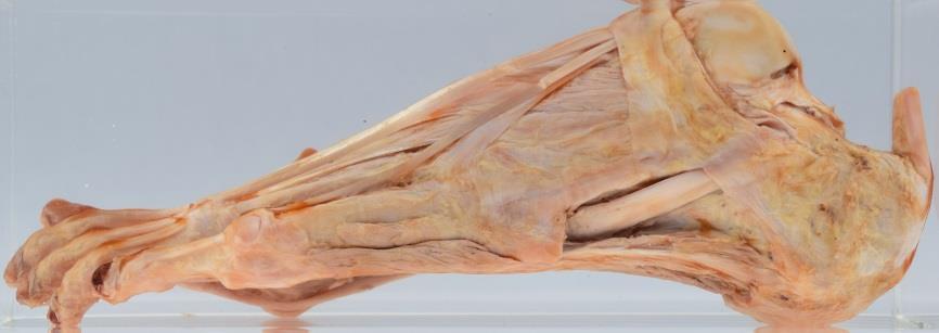
5 A Lateral view of the left foot 1. Abductor hallucis 2. Flexor digitorum brevis 屈趾短肌 3. Abductor digiti minimi 外展小趾肌 4. Flexor digiti minimi brevis 屈小趾短肌 5. 2 nd lumbrical 第二蚓狀肌 6. 1 st lumbrical 第一蚓狀肌 7. Flexor hallucis (lateral head) ( 外側頭 ) 8. Flexor hallucis (medial head) ( 內側頭 ) 9. Plantar aponeurosis 足底腱膜 10. Medial cuneiform 內側楔狀骨 st metatarsal 第一蹠骨 12. Flexor hallucis longus tendon 13. Flexor digitorum brevis tendons 屈趾短肌肌腱 14. Flexor digitorum longus tendons 屈趾長肌肌腱. Fibularis longus tendon 腓骨長肌肌腱. Extensor digitorum longus tendon 伸趾長肌腱 17. Fibularis tertius tendon 第三腓骨肌腱 18. Fibularis brevis tendon 腓骨短肌腱 19. Inferior fibular retinaculum 下腓骨肌支持帶 20. Tuberosity of 5 th metatarsal 第五蹠骨粗隆 21. Dorsal aponeurosis 背側腱膜 22. Extensor digitorum brevis 伸趾短肌 th metatarsal 第五蹠骨 th proximal phalanx 第五近側趾骨 25. Calcaneus 跟骨 26. Flexor digitorum longus tendon 屈趾長肌肌腱 27. Extensor hallucis longus tendon 28. Extensor hallucis brevis 29. Tibialis anterior tendon 脛前肌肌腱 30. Tibia 脛骨 31. Fibula 腓骨
 ( 外側頭 ) 8. Flexor hallucis (medial head) ( 內側頭 ) 9. Plantar aponeurosis 足底腱膜 10. Medial cuneiform 內側楔狀骨 11.")
6 A Medial view of the left foot 1. Abductor hallucis 2. Flexor digitorum brevis 屈趾短肌 3. Abductor digiti minimi 外展小趾肌 4. Flexor digiti minimi brevis 屈小趾短肌 5. 2 nd lumbrical 第二蚓狀肌 6. 1 st lumbrical 第一蚓狀肌 7. Flexor hallucis (lateral head) ( 外側頭 ) 8. Flexor hallucis (medial head) ( 內側頭 ) 9. Plantar aponeurosis 足底腱膜 10. Medial cuneiform 內側楔狀骨 st metatarsal 第一蹠骨 12. Flexor hallucis longus tendon 13. Flexor digitorum brevis tendons 屈趾短肌肌腱 14. Flexor digitorum longus tendons 屈趾長肌肌腱. Fibularis longus tendon 腓骨長肌肌腱. Extensor digitorum longus tendon 伸趾長肌腱 17. Fibularis tertius tendon 第三腓骨肌腱 18. Fibularis brevis tendon 腓骨短肌腱 19. Inferior fibular retinaculum 下腓骨肌支持帶 20. Tuberosity of 5 th metatarsal 第五蹠骨粗隆 21. Dorsal aponeurosis 背側腱膜 22. Extensor digitorum brevis 伸趾短肌 th metatarsal 第五蹠骨 th proximal phalanx 第五近側趾骨 25. Calcaneus 跟骨 26. Flexor digitorum longus tendon 屈趾長肌肌腱 27. Extensor hallucis longus tendon 28. Extensor hallucis brevis 29. Tibialis anterior tendon 脛前肌肌腱 30. Tibia 脛骨 31. Fibula 腓骨
 ( 外側頭 ) 8. Flexor hallucis (medial head) ( 內側頭 ) 9. Plantar aponeurosis 足底腱膜 10.")
7 18 A Plantar view of the left foot 1. Abductor hallucis 2. Flexor digitorum brevis 屈趾短肌 3. Abductor digiti minimi 外展小趾肌 4. Flexor digiti minimi brevis 屈小趾短肌 5. 2 nd lumbrical 第二蚓狀肌 6. 1 st lumbrical 第一蚓狀肌 7. Flexor hallucis (lateral head) ( 外側頭 ) 8. Flexor hallucis (medial head) ( 內側頭 ) 9. Plantar aponeurosis 足底腱膜 10. Medial cuneiform 內側楔狀骨 st metatarsal 第一蹠骨 12. Flexor hallucis longus tendon 13. Flexor digitorum brevis tendons 屈趾短肌肌腱 14. Flexor digitorum longus tendons 屈趾長肌肌腱. Fibularis longus tendon 腓骨長肌肌腱. Extensor digitorum longus tendon 伸趾長肌腱 17. Fibularis tertius tendon 第三腓骨肌腱 18. Fibularis brevis tendon 腓骨短肌腱 19. Inferior fibular retinaculum 下腓骨肌支持帶 20. Tuberosity of 5 th metatarsal 第五蹠骨粗隆 21. Dorsal aponeurosis 背側腱膜 22. Extensor digitorum brevis 伸趾短肌 th metatarsal 第五蹠骨 th proximal phalanx 第五近側趾骨 25. Calcaneus 跟骨 26. Flexor digitorum longus tendon 屈趾長肌肌腱 27. Extensor hallucis longus tendon 28. Extensor hallucis brevis 29. Tibialis anterior tendon 脛前肌肌腱 30. Tibia 脛骨 31. Fibula 腓骨
 ( 外側頭 ) 8. Flexor hallucis (medial head) ( 內側頭 ) 9. Plantar aponeurosis 足底腱膜 10. Medial cuneiform 內側楔狀骨 11.")
8 31 A Dorsal view of the left foot 1. Abductor hallucis 2. Flexor digitorum brevis 屈趾短肌 3. Abductor digiti minimi 外展小趾肌 4. Flexor digiti minimi brevis 屈小趾短肌 5. 2 nd lumbrical 第二蚓狀肌 6. 1 st lumbrical 第一蚓狀肌 7. Flexor hallucis (lateral head) ( 外側頭 ) 8. Flexor hallucis (medial head) ( 內側頭 ) 9. Plantar aponeurosis 足底腱膜 10. Medial cuneiform 內側楔狀骨 st metatarsal 第一蹠骨 12. Flexor hallucis longus tendon 13. Flexor digitorum brevis tendons 屈趾短肌肌腱 14. Flexor digitorum longus tendons 屈趾長肌肌腱. Fibularis longus tendon 腓骨長肌肌腱. Extensor digitorum longus tendon 伸趾長肌腱 17. Fibularis tertius tendon 第三腓骨肌腱 18. Fibularis brevis tendon 腓骨短肌腱 19. Inferior fibular retinaculum 下腓骨肌支持帶 20. Tuberosity of 5 th metatarsal 第五蹠骨粗隆 21. Dorsal aponeurosis 背側腱膜 22. Extensor digitorum brevis 伸趾短肌 th metatarsal 第五蹠骨 th proximal phalanx 第五近側趾骨 25. Calcaneus 跟骨 26. Flexor digitorum longus tendon 屈趾長肌肌腱 27. Extensor hallucis longus tendon 28. Extensor hallucis brevis 29. Tibialis anterior tendon 脛前肌肌腱 30. Tibia 脛骨 31. Fibula 腓骨

9 A09-1 Posterior view of the left foot Extensor digitorum longus tendon 伸趾長肌腱 27. Extensor hallucis longus tendon 28. Extensor hallucis brevis 29. Tibialis anterior tendon 脛前肌肌腱 30. Tibia 脛骨 31. Fibula 腓骨 32. Calcaneal tendon 跟腱 32

10 A09-1 Anterior view of the right foot Extensor digitorum longus tendon 伸趾長肌腱 27. Extensor hallucis longus tendon 28. Extensor hallucis brevis 29. Tibialis anterior tendon 脛前肌肌腱
11 A09-2
12 A09-2 Lateral view of the left foot Plantar aponeurosis 足底腱膜 2. Flexor digitorum brevis 屈趾短肌 3. Quadratus plante 蹠方肌 4. Abductor hallucis 5. Flexor hallucis brevis 6. Flexor digitorum longus tendons 屈趾長肌肌腱 7. Flexor hallucis longus tendon 8. 1 st lumbrical 第一蚓狀肌 9. 2 nd lumbrical 第二蚓狀肌 rd lumbrical 第三蚓狀肌 th lumbrical 第四蚓狀肌 12. Abductor digiti minimi 外展小趾肌 13. Flexor digiti minimi brevis 屈小趾短肌 14. Fibularis tertius 第三腓骨肌. Extensor digitorum longus tendons 伸趾長肌腱. Extensor digitorum brevis 伸趾短肌 17. Inferior fibular retinaculum 下腓骨肌支持帶 18. Fibularis brevis tendon 腓骨短肌腱 19. Fibularis longus tendon 腓骨長肌肌腱 20. Long plantar ligament 足底長韌帶 21. Calcaneal ligament 跟腱 22. Calcaneus 跟骨. 23. Talus 距骨 24. Tibialis anterior 脛前肌 25. Extensor hallucis longus tendon 26. Inferior extensor retinaculum 下伸肌支持帶 27. Flexor digitorum brevis tendon 屈趾短肌肌腱 28. Extensor hallucis brevis

13 Medial view of the left foot A Plantar aponeurosis 足底腱膜 2. Flexor digitorum brevis 屈趾短肌 3. Quadratus plante 蹠方肌 4. Abductor hallucis 5. Flexor hallucis brevis 6. Flexor digitorum longus tendons 屈趾長肌肌腱 7. Flexor hallucis longus tendon 8. 1 st lumbrical 第一蚓狀肌 9. 2 nd lumbrical 第二蚓狀肌 rd lumbrical 第三蚓狀肌 th lumbrical 第四蚓狀肌 12. Abductor digiti minimi 外展小趾肌 13. Flexor digiti minimi brevis 屈小趾短肌 14. Fibularis tertius 第三腓骨肌. Extensor digitorum longus tendons 伸趾長肌腱. Extensor digitorum brevis 伸趾短肌 17. Inferior fibular retinaculum 下腓骨肌支持帶 18. Fibularis brevis tendon 腓骨短肌腱 19. Fibularis longus tendon 腓骨長肌肌腱 20. Long plantar ligament 足底長韌帶 21. Calcaneal ligament 跟腱 22. Calcaneus 跟骨. 23. Talus 距骨 24. Tibialis anterior 脛前肌 25. Extensor hallucis longus tendon 26. Inferior extensor retinaculum 下伸肌支持帶 27. Flexor digitorum brevis tendon 屈趾短肌肌腱 28. Extensor hallucis brevis

14 Plantar view of the left foot A Plantar aponeurosis 足底腱膜 2. Flexor digitorum brevis 屈趾短肌 3. Quadratus plante 蹠方肌 4. Abductor hallucis 5. Flexor hallucis brevis 6. Flexor digitorum longus tendons 屈趾長肌肌腱 7. Flexor hallucis longus tendon 8. 1 st lumbrical 第一蚓狀肌 9. 2 nd lumbrical 第二蚓狀肌 rd lumbrical 第三蚓狀肌 th lumbrical 第四蚓狀肌 12. Abductor digiti minimi 外展小趾肌 13. Flexor digiti minimi brevis 屈小趾短肌 14. Fibularis tertius 第三腓骨肌. Extensor digitorum longus tendons 伸趾長肌腱. Extensor digitorum brevis 伸趾短肌 17. Inferior fibular retinaculum 下腓骨肌支持帶 18. Fibularis brevis tendon 腓骨短肌腱 19. Fibularis longus tendon 腓骨長肌肌腱 20. Long plantar ligament 足底長韌帶 21. Calcaneal ligament 跟腱 22. Calcaneus 跟骨. 23. Talus 距骨 24. Tibialis anterior 脛前肌 25. Extensor hallucis longus tendon 26. Inferior extensor retinaculum 下伸肌支持帶 27. Flexor digitorum brevis tendon 屈趾短肌肌腱 28. Extensor hallucis brevis

15 Dorsal view of the left foot A Plantar aponeurosis 足底腱膜 2. Flexor digitorum brevis 屈趾短肌 3. Quadratus plante 蹠方肌 4. Abductor hallucis 5. Flexor hallucis brevis 6. Flexor digitorum longus tendons 屈趾長肌肌腱 7. Flexor hallucis longus tendon 8. 1 st lumbrical 第一蚓狀肌 9. 2 nd lumbrical 第二蚓狀肌 rd lumbrical 第三蚓狀肌 th lumbrical 第四蚓狀肌 12. Abductor digiti minimi 外展小趾肌 13. Flexor digiti minimi brevis 屈小趾短肌 14. Fibularis tertius 第三腓骨肌. Extensor digitorum longus tendons 伸趾長肌腱. Extensor digitorum brevis 伸趾短肌 17. Inferior fibular retinaculum 下腓骨肌支持帶 18. Fibularis brevis tendon 腓骨短肌腱 19. Fibularis longus tendon 腓骨長肌肌腱 20. Long plantar ligament 足底長韌帶 21. Calcaneal ligament 跟腱 22. Calcaneus 跟骨. 23. Talus 距骨 24. Tibialis anterior 脛前肌 25. Extensor hallucis longus tendon 26. Inferior extensor retinaculum 下伸肌支持帶 27. Flexor digitorum brevis tendon 屈趾短肌肌腱 28. Extensor hallucis brevis

16 A09-2 Posterior view of the left foot Extensor digitorum brevis 伸趾短肌 17. Inferior fibular retinaculum 下腓骨肌支持帶 18. Fibularis brevis tendon 腓骨短肌腱 19. Fibularis longus tendon 腓骨長肌肌腱 20. Long plantar ligament 足底長韌帶 21. Calcaneal ligament 跟腱 22. Calcaneus 跟骨. 23. Talus 距骨 24. Tibialis anterior 脛前肌 25. Extensor hallucis longus tendon 26. Inferior extensor retinaculum 下伸肌支持帶 27. Flexor digitorum brevis tendon 屈趾短肌肌腱 28. Extensor hallucis brevis 29. Tibialis posterior 脛後肌 22

17 A Anterior view of the left foot Fibularis tertius tendon 第三腓骨肌肌腱. Extensor digitorum longus tendons 伸趾長肌腱. Extensor digitorum brevis 伸趾短肌 17. Inferior fibular retinaculum 下腓骨肌支持帶 24. Tibialis anterior 脛前肌 25. Extensor hallucis longus tendon 26. Inferior extensor retinaculum 下伸肌支持帶 27. Flexor digitorum brevis tendon 屈趾短肌肌腱 28. Extensor hallucis brevis
18 A09-3

19 1. Flexor digitorum brevis 屈趾短肌 2. Quadratus plante 蹠方肌 3. Abductor hallucis 4. Flexor hallucis brevis 5. Adductor hallucis (oblique head) ( 斜頭 ) 6. Plantar interossei 掌骨間肌 7. Flexor digiti minimi brevis 屈小趾短肌 8. Flexor digitorum longus tendons 屈趾長肌肌腱 9. Lumbrical 蚓狀肌 10. Dorsal interossei 足背骨間肌 11. Extensor digitorum brevis 伸趾短肌 12. Extensor hallucis brevis 13. Extensor hallucis longus tendon 14. Tibialis anterior 脛前肌. Extensor digitorum longus tendons 伸趾長肌腱. Fibularis tertius tendon 第三腓骨肌肌腱 Medial view of the right foot A Inferior extensor retinaculum 下伸肌支持帶 18. Superior extensor retinaculum 上伸肌支持帶 19. Fibula 腓骨 20. Tibia 脛骨 21. Fibularis brevis tendon 腓骨短肌腱 22. Fibularis longus tendon 腓骨長肌肌腱 23. Calcaneal ligament 跟腱 24. Flexor hallucis longus tendon 25. Plantar aponeurosis 掌腱膜 26. Abductor digiti minimi 小趾外展肌 27. Calcaneus 跟骨
20 A Lateral view of the right foot Flexor digitorum brevis 屈趾短肌 2. Quadratus plante 蹠方肌 3. Abductor hallucis 4. Flexor hallucis brevis 5. Adductor hallucis (oblique head) ( 斜頭 ) 6. Plantar interossei 掌骨間肌 7. Flexor digiti minimi brevis 屈小趾短肌 8. Flexor digitorum longus tendons 屈趾長肌肌腱 9. Lumbrical 蚓狀肌 10. Dorsal interossei 足背骨間肌 11. Extensor digitorum brevis 伸趾短肌 12. Extensor hallucis brevis 13. Extensor hallucis longus tendon Tibialis anterior 脛前肌. Extensor digitorum longus tendons 伸趾長肌腱. Fibularis tertius tendon 第三腓骨肌肌腱 17. Inferior extensor retinaculum 下伸肌支持帶 18. Superior extensor retinaculum 上伸肌支持帶 19. Fibula 腓骨 20. Tibia 脛骨 21. Fibularis brevis tendon 腓骨短肌腱 22. Fibularis longus tendon 腓骨長肌肌腱 23. Calcaneal ligament 跟腱 24. Flexor hallucis longus tendon 25. Plantar aponeurosis 掌腱膜 26. Abductor digiti minimi 小趾外展肌 27. Calcaneus 跟骨
 ( 斜頭 ) 6. Plantar interossei 掌骨間肌 7. Flexor digiti minimi brevis 屈小趾短肌 8. Flexor digitorum longus tendons 屈趾長肌肌腱 9. Lumbrical 蚓狀肌 10. Dorsal interossei 足背骨間肌 11.")
21 3 A Plantar view of the right foot Flexor digitorum brevis 屈趾短肌 2. Quadratus plante 蹠方肌 3. Abductor hallucis 4. Flexor hallucis brevis 5. Adductor hallucis (oblique head) ( 斜頭 ) 6. Plantar interossei 掌骨間肌 7. Flexor digiti minimi brevis 屈小趾短肌 8. Flexor digitorum longus tendons 屈趾長肌肌腱 9. Lumbrical 蚓狀肌 10. Dorsal interossei 足背骨間肌 11. Extensor digitorum brevis 伸趾短肌 12. Extensor hallucis brevis 13. Extensor hallucis longus tendon 14. Tibialis anterior 脛前肌. Extensor digitorum longus tendons 伸趾長肌腱. Fibularis tertius tendon 第三腓骨肌肌腱 17. Inferior extensor retinaculum 下伸肌支持帶 18. Superior extensor retinaculum 上伸肌支持帶 19. Fibula 腓骨 20. Tibia 脛骨 21. Fibularis brevis tendon 腓骨短肌腱 22. Fibularis longus tendon 腓骨長肌肌腱 23. Calcaneal ligament 跟腱 24. Flexor hallucis longus tendon 25. Plantar aponeurosis 掌腱膜 26. Abductor digiti minimi 小趾外展肌 27. Calcaneus 跟骨
 ( 斜頭 ) 6. Plantar interossei 掌骨間肌 7. Flexor digiti minimi brevis 屈小趾短肌 8. Flexor digitorum longus tendons 屈趾長肌肌腱 9. Lumbrical 蚓狀肌 10. Dorsal interossei 足背骨間肌 11.")
22 10 A Dorsal view of the right foot 1. Flexor digitorum brevis 屈趾短肌 2. Quadratus plante 蹠方肌 3. Abductor hallucis 4. Flexor hallucis brevis 5. Adductor hallucis (oblique head) ( 斜頭 ) 6. Plantar interossei 掌骨間肌 7. Flexor digiti minimi brevis 屈小趾短肌 8. Flexor digitorum longus tendons 屈趾長肌肌腱 9. Lumbrical 蚓狀肌 10. Dorsal interossei 足背骨間肌 11. Extensor digitorum brevis 伸趾短肌 12. Extensor hallucis brevis 13. Extensor hallucis longus tendon 14. Tibialis anterior 脛前肌. Extensor digitorum longus tendons 伸趾長肌腱. Fibularis tertius tendon 第三腓骨肌肌腱 17. Inferior extensor retinaculum 下伸肌支持帶 18. Superior extensor retinaculum 上伸肌支持帶 19. Fibula 腓骨 20. Tibia 脛骨 21. Fibularis brevis tendon 腓骨短肌腱 22. Fibularis longus tendon 腓骨長肌肌腱 23. Calcaneal ligament 跟腱 24. Flexor hallucis longus tendon 25. Plantar aponeurosis 掌腱膜 26. Abductor digiti minimi 小趾外展肌 27. Calcaneus 跟骨
 ( 斜頭 ) 6. Plantar interossei 掌骨間肌 7. Flexor digiti minimi brevis 屈小趾短肌 8. Flexor digitorum longus tendons 屈趾長肌肌腱 9. Lumbrical 蚓狀肌 10. Dorsal interossei 足背骨間肌 11.")
23 20 19 A09-3 Posterior view of the right foot Flexor digitorum brevis 屈趾短肌 2. Quadratus plante 蹠方肌 3. Abductor hallucis 4. Flexor hallucis brevis 5. Adductor hallucis (oblique head) ( 斜頭 ) 6. Plantar interossei 掌骨間肌 7. Flexor digiti minimi brevis 屈小趾短肌 8. Flexor digitorum longus tendons 屈趾長肌肌腱 9. Lumbrical 蚓狀肌 10. Dorsal interossei 足背骨間肌 11. Extensor digitorum brevis 伸趾短肌 12. Extensor hallucis brevis 13. Extensor hallucis longus tendon 14. Tibialis anterior 脛前肌. Extensor digitorum longus tendons 伸趾長肌腱. Fibularis tertius tendon 第三腓骨肌肌腱 17. Inferior extensor retinaculum 下伸肌支持帶 18. Superior extensor retinaculum 上伸肌支持帶 19. Fibula 腓骨 20. Tibia 脛骨 21. Fibularis brevis tendon 腓骨短肌腱 22. Fibularis longus tendon 腓骨長肌肌腱 23. Calcaneal ligament 跟腱 24. Flexor hallucis longus tendon 25. Plantar aponeurosis 掌腱膜 26. Abductor digiti minimi 小趾外展肌 27. Calcaneus 跟骨
 ( 斜頭 ) 6. Plantar interossei 掌骨間肌 7. Flexor digiti minimi brevis 屈小趾短肌 8. Flexor digitorum longus tendons 屈趾長肌肌腱 9. Lumbrical 蚓狀肌 10. Dorsal interossei 足背骨間肌 11.")
24 A Anterior view of the right foot Flexor digitorum brevis 屈趾短肌 2. Quadratus plante 蹠方肌 3. Abductor hallucis 4. Flexor hallucis brevis 5. Adductor hallucis (oblique head) ( 斜頭 ) 6. Plantar interossei 掌骨間肌 7. Flexor digiti minimi brevis 屈小趾短肌 8. Flexor digitorum longus tendons 屈趾長肌肌腱 9. Lumbrical 蚓狀肌 10. Dorsal interossei 足背骨間肌 11. Extensor digitorum brevis 伸趾短肌 12. Extensor hallucis brevis 13. Extensor hallucis longus tendon 14. Tibialis anterior 脛前肌. Extensor digitorum longus tendons 伸趾長肌腱. Fibularis tertius tendon 第三腓骨肌肌腱 17. Inferior extensor retinaculum 下伸肌支持帶 18. Superior extensor retinaculum 上伸肌支持帶 19. Fibula 腓骨 20. Tibia 脛骨 21. Fibularis brevis tendon 腓骨短肌腱 22. Fibularis longus tendon 腓骨長肌肌腱 23. Calcaneal ligament 跟腱 24. Flexor hallucis longus tendon 25. Plantar aponeurosis 掌腱膜 26. Abductor digiti minimi 小趾外展肌 27. Calcaneus 跟骨
25 A09-4

26 1. Long plantar ligament 足底長韌帶 2. Adductor hallucis (oblique head) ( 斜頭 ) 3. Adductor hallucis (transverse head) ( 橫頭 ) 4. Flexor digiti minimi brevis 屈小趾短肌 5. Plantar interossei 足底骨間肌 6. Flexor digitorum brevis 屈趾短肌 7. Interossei 骨間肌 8. Fibularis longus tendon 腓骨長肌肌腱 9. Fibularis brevis tendon 腓骨短肌腱 10. Flexor hallucis brevis 11. Inferior extensor retinaculum 下伸肌支持帶 Lateral view of the left foot A Superior extensor retinaculum 上伸肌支持帶 13. Calcaneus tendon 跟腱 14. Tibialis anterior 脛前肌. Extensor hallucis longus tendon. Extensor digitorum longus tendons 伸趾長肌腱 17. Fibula 腓骨 18. Tibia 脛骨 19. Calcaneus 跟骨 20. Cuboid 骰骨 21. Navicular 舟狀骨 22. Tibialis posterior 脛後肌 23. Flexor digitorum longus tendon (groove) 屈趾長肌肌腱 ( 溝 ) 24. Flexor hallucis longus tendon (groove) ( 溝 ) 25. Flexor retinaculum 屈肌支持帶 26. Posterior tibial artery 脛後動脈 27. Anterior tibial artery 脛前動脈
27 A Medial view of the left foot Long plantar ligament 足底長韌帶 2. Adductor hallucis (oblique head) ( 斜頭 ) 3. Adductor hallucis (transverse head) ( 橫頭 ) 4. Flexor digiti minimi brevis 屈小趾短肌 5. Plantar interossei 足底骨間肌 6. Flexor digitorum brevis 屈趾短肌 7. Interossei 骨間肌 8. Fibularis longus tendon 腓骨長肌肌腱 9. Fibularis brevis tendon 腓骨短肌腱 10. Flexor hallucis brevis 11. Inferior extensor retinaculum 下伸肌支持帶 Superior extensor retinaculum 上伸肌支持帶 13. Calcaneus tendon 跟腱 14. Tibialis anterior 脛前肌. Extensor hallucis longus tendon. Extensor digitorum longus tendons 伸趾長肌腱 17. Fibula 腓骨 18. Tibia 脛骨 19. Calcaneus 跟骨 20. Cuboid 骰骨 21. Navicular 舟狀骨 22. Tibialis posterior 脛後肌 23. Flexor digitorum longus tendon (groove) 屈趾長肌肌腱 ( 溝 ) 24. Flexor hallucis longus tendon (groove) ( 溝 ) 25. Flexor retinaculum 屈肌支持帶 26. Posterior tibial artery 脛後動脈 27. Anterior tibial artery 脛前動脈

28 A Plantar view of the left foot 1. Long plantar ligament 足底長韌帶 2. Adductor hallucis (oblique head) ( 斜頭 ) 3. Adductor hallucis (transverse head) ( 橫頭 ) 4. Flexor digiti minimi brevis 屈小趾短肌 5. Plantar interossei 足底骨間肌 6. Flexor digitorum brevis 屈趾短肌 7. Interossei 骨間肌 8. Fibularis longus tendon 腓骨長肌肌腱 9. Fibularis brevis tendon 腓骨短肌腱 10. Flexor hallucis brevis 11. Inferior extensor retinaculum 下伸肌支持帶 12. Superior extensor retinaculum 上伸肌支持帶 13. Calcaneus tendon 跟腱 14. Tibialis anterior 脛前肌. Extensor hallucis longus tendon. Extensor digitorum longus tendons 伸趾長肌腱 17. Fibula 腓骨 18. Tibia 脛骨 19. Calcaneus 跟骨 20. Cuboid 骰骨 21. Navicular 舟狀骨 22. Tibialis posterior 脛後肌 23. Flexor digitorum longus tendon (groove) 屈趾長肌肌腱 ( 溝 ) 24. Flexor hallucis longus tendon (groove) ( 溝 ) 25. Flexor retinaculum 屈肌支持帶 26. Posterior tibial artery 脛後動脈 27. Anterior tibial artery 脛前動脈

29 A Dorsal view of the left foot 1. Long plantar ligament 足底長韌帶 2. Adductor hallucis (oblique head) ( 斜頭 ) 3. Adductor hallucis (transverse head) ( 橫頭 ) 4. Flexor digiti minimi brevis 屈小趾短肌 5. Plantar interossei 足底骨間肌 6. Flexor digitorum brevis 屈趾短肌 7. Interossei 骨間肌 8. Fibularis longus tendon 腓骨長肌肌腱 9. Fibularis brevis tendon 腓骨短肌腱 10. Flexor hallucis brevis 11. Inferior extensor retinaculum 下伸肌支持帶 12. Superior extensor retinaculum 上伸肌支持帶 13. Calcaneus tendon 跟腱 14. Tibialis anterior 脛前肌. Extensor hallucis longus tendon. Extensor digitorum longus tendons 伸趾長肌腱 17. Fibula 腓骨 18. Tibia 脛骨 19. Calcaneus 跟骨 20. Cuboid 骰骨 21. Navicular 舟狀骨 22. Tibialis posterior 脛後肌 23. Flexor digitorum longus tendon 屈趾長肌肌腱 24. Flexor hallucis longus tendon (groove) ( 溝 ) 25. Flexor retinaculum 屈肌支持帶 26. Posterior tibial artery 脛後動脈 27. Anterior tibial artery 脛前動脈

30 A Posterior view of the left foot 1. Long plantar ligament 足底長韌帶 2. Adductor hallucis (oblique head) ( 斜頭 ) 3. Adductor hallucis (transverse head) ( 橫頭 ) 4. Flexor digiti minimi brevis 屈小趾短肌 5. Plantar interossei 足底骨間肌 6. Flexor digitorum brevis 屈趾短肌 7. Interossei 骨間肌 8. Fibularis longus tendon 腓骨長肌肌腱 9. Fibularis brevis tendon 腓骨短肌腱 10. Flexor hallucis brevis 11. Inferior extensor retinaculum 下伸肌支持帶 12. Superior extensor retinaculum 上伸肌支持帶 13. Calcaneus tendon 跟腱 14. Tibialis anterior 脛前肌. Extensor hallucis longus tendon. Extensor digitorum longus tendons 伸趾長肌腱 17. Fibula 腓骨 18. Tibia 脛骨 19. Calcaneus 跟骨 20. Cuboid 骰骨 21. Navicular 舟狀骨 22. Tibialis posterior 脛後肌 23. Flexor digitorum longus tendon 屈趾長肌肌腱 24. Flexor hallucis longus tendon 25. Flexor retinaculum 屈肌支持帶 26. Posterior tibial artery 脛後動脈 27. Anterior tibial artery 脛前動脈


31 A Anterior view of the left foot Long plantar ligament 足底長韌帶 2. Adductor hallucis (oblique head) ( 斜頭 ) 3. Adductor hallucis (transverse head) ( 橫頭 ) 4. Flexor digiti minimi brevis 屈小趾短肌 5. Plantar interossei 足底骨間肌 6. Flexor digitorum brevis 屈趾短肌 7. Interossei 骨間肌 8. Fibularis longus tendon 腓骨長肌肌腱 9. Fibularis brevis tendon 腓骨短肌腱 10. Flexor hallucis brevis 11. Inferior extensor retinaculum 下伸肌支持帶 12. Superior extensor retinaculum 上伸肌支持帶 13. Calcaneus tendon 跟腱 14. Tibialis anterior 脛前肌. Extensor hallucis longus tendon. Extensor digitorum longus tendons 伸趾長肌腱 17. Fibula 腓骨 18. Tibia 脛骨 19. Calcaneus 跟骨 20. Cuboid 骰骨 21. Navicular 舟狀骨 22. Tibialis posterior 脛後肌 23. Flexor digitorum longus tendon (groove) 屈趾長肌肌腱 ( 溝 ) 24. Flexor hallucis longus tendon (groove) 腱 ( 溝 ) 25. Flexor retinaculum 屈肌支持帶 26. Posterior tibial artery 脛後動脈 27. Anterior tibial artery 脛前動脈
32 A09-5
 跟舟韌帶 13. Fibularis longus tendon (groove) 腓骨長肌肌腱 ( 溝 ) 14.")
33 A09-5 Medial view of the right foot Talus 距骨 2. Calcaneus 跟骨 3. Navicular 舟狀骨 4. Medial cuneiform 內側楔狀骨 5. Intermediate cuneiform 中間楔狀骨 6. Lateral cuneiform 外側楔狀骨 7. Cuboid 骰骨 8. Tibia 脛骨 9. Fibula 腓骨 10. Calcaneus tendon 跟腱 11. Long plantar ligament 足底長韌帶 12. Plantar calcaneonavicular ligament 足底 ( 蹠 ) 跟舟韌帶 13. Fibularis longus tendon (groove) 腓骨長肌肌腱 ( 溝 ) 14. Anterior talofibular ligament 前距腓韌帶. Interosseous talocalcaneus ligament 距跟骨間韌帶. Flexor hallucis longus tendon (groove) ( 溝 ) 17. Flexor digitorum longus tendon (groove) 屈趾長肌肌腱 ( 溝 ) 18. Tibialis posterior tendon (groove) 脛後肌肌腱 ( 溝 ) 19. Fibularis brevis tendon (groove) 腓骨短肌肌腱 ( 溝 )
 跟舟韌帶 13. Fibularis longus tendon (groove) 腓骨長肌肌腱 ( 溝 ) 14.")
34 8 Lateral view of the right foot A Talus 距骨 2. Calcaneus 跟骨 3. Navicular 舟狀骨 4. Medial cuneiform 內側楔狀骨 5. Intermediate cuneiform 中間楔狀骨 6. Lateral cuneiform 外側楔狀骨 7. Cuboid 骰骨 8. Tibia 脛骨 9. Fibula 腓骨 10. Calcaneus tendon 跟腱 11. Long plantar ligament 足底長韌帶 12. Plantar calcaneonavicular ligament 足底 ( 蹠 ) 跟舟韌帶 13. Fibularis longus tendon (groove) 腓骨長肌肌腱 ( 溝 ) 14. Anterior talofibular ligament 前距腓韌帶. Interosseous talocalcaneus ligament 距跟骨間韌帶. Flexor hallucis longus tendon (groove) ( 溝 ) 17. Flexor digitorum longus tendon (groove) 屈趾長肌肌腱 ( 溝 ) 18. Tibialis posterior tendon (groove) 脛後肌肌腱 ( 溝 ) 19. Fibularis brevis tendon (groove) 腓骨短肌肌腱 ( 溝 )
 跟舟韌帶 13. Fibularis longus tendon (groove) 腓骨長肌肌腱 ( 溝 ) 14.")
35 A Plantar view of the right foot 1. Talus 距骨 2. Calcaneus 跟骨 3. Navicular 舟狀骨 4. Medial cuneiform 內側楔狀骨 5. Intermediate cuneiform 中間楔狀骨 6. Lateral cuneiform 外側楔狀骨 7. Cuboid 骰骨 8. Tibia 脛骨 9. Fibula 腓骨 10. Calcaneus tendon 跟腱 11. Long plantar ligament 足底長韌帶 12. Plantar calcaneonavicular ligament 足底 ( 蹠 ) 跟舟韌帶 13. Fibularis longus tendon (groove) 腓骨長肌肌腱 ( 溝 ) 14. Anterior talofibular ligament 前距腓韌帶. Interosseous talocalcaneus ligament 距跟骨間韌帶. Flexor hallucis longus tendon (groove) ( 溝 ) 17. Flexor digitorum longus tendon (groove) 屈趾長肌肌腱 ( 溝 ) 18. Tibialis posterior tendon (groove) 脛後肌肌腱 ( 溝 ) 19. Fibularis brevis tendon (groove) 腓骨短肌肌腱 ( 溝 )
 跟舟韌帶 13. Fibularis longus tendon (groove) 腓骨長肌肌腱 ( 溝 ) 14.")
36 A Dorsal view of the right foot 1. Talus 距骨 2. Calcaneus 跟骨 3. Navicular 舟狀骨 4. Medial cuneiform 內側楔狀骨 5. Intermediate cuneiform 中間楔狀骨 6. Lateral cuneiform 外側楔狀骨 7. Cuboid 骰骨 8. Tibia 脛骨 9. Fibula 腓骨 10. Calcaneus tendon 跟腱 11. Long plantar ligament 足底長韌帶 12. Plantar calcaneonavicular ligament 足底 ( 蹠 ) 跟舟韌帶 13. Fibularis longus tendon (groove) 腓骨長肌肌腱 ( 溝 ) 14. Anterior talofibular ligament 前距腓韌帶. Interosseous talocalcaneus ligament 距跟骨間韌帶. Flexor hallucis longus tendon (groove) ( 溝 ) 17. Flexor digitorum longus tendon (groove) 屈趾長肌肌腱 ( 溝 ) 18. Tibialis posterior tendon (groove) 脛後肌肌腱 ( 溝 ) 19. Fibularis brevis tendon (groove) 腓骨短肌肌腱 ( 溝 )
 跟舟韌帶 13. Fibularis longus tendon (groove) 腓骨長肌肌腱 ( 溝 ) 14.")
37 A Posterior view of the right foot 1. Talus 距骨 2. Calcaneus 跟骨 3. Navicular 舟狀骨 4. Medial cuneiform 內側楔狀骨 5. Intermediate cuneiform 中間楔狀骨 6. Lateral cuneiform 外側楔狀骨 7. Cuboid 骰骨 8. Tibia 脛骨 9. Fibula 腓骨 10. Calcaneus tendon 跟腱 11. Long plantar ligament 足底長韌帶 12. Plantar calcaneonavicular ligament 足底 ( 蹠 ) 跟舟韌帶 13. Fibularis longus tendon (groove) 腓骨長肌肌腱 ( 溝 ) 14. Anterior talofibular ligament 前距腓韌帶. Interosseous talocalcaneus ligament 距跟骨間韌帶. Flexor hallucis longus tendon (groove) ( 溝 ) 17. Flexor digitorum longus tendon (groove) 屈趾長肌肌腱 ( 溝 ) 18. Tibialis posterior tendon (groove) 脛後肌肌腱 ( 溝 ) 19. Fibularis brevis tendon (groove) 腓骨短肌肌腱 ( 溝 ) 2
 跟舟韌帶 13. Fibularis longus tendon (groove) 腓骨長肌肌腱 ( 溝 ) 14.")
38 A09-5 Anterior view of the right foot Talus 距骨 2. Calcaneus 跟骨 3. Navicular 舟狀骨 4. Medial cuneiform 內側楔狀骨 5. Intermediate cuneiform 中間楔狀骨 6. Lateral cuneiform 外側楔狀骨 7. Cuboid 骰骨 8. Tibia 脛骨 9. Fibula 腓骨 10. Calcaneus tendon 跟腱 11. Long plantar ligament 足底長韌帶 12. Plantar calcaneonavicular ligament 足底 ( 蹠 ) 跟舟韌帶 13. Fibularis longus tendon (groove) 腓骨長肌肌腱 ( 溝 ) 14. Anterior talofibular ligament 前距腓韌帶. Interosseous talocalcaneus ligament 距跟骨間韌帶. Flexor hallucis longus tendon (groove) ( 溝 ) 17. Flexor digitorum longus tendon (groove) 屈趾長肌肌腱 ( 溝 ) 18. Tibialis posterior tendon (groove) 脛後肌肌腱 ( 溝 ) 19. Fibularis brevis tendon (groove) 腓骨短肌肌腱 ( 溝 )
39 A10-3
 collateral ligament 外側副韌帶 9. Medial (tibial) collateral ligament 內側副韌帶 10. Patella 髕骨 ( 後表面 ) 11. Lateral condyle of tibia 外側脛髁 12.")
40 A10-3 Anterior view of the left knee Femur 股骨 2. Medial condyle of femur 內側股髁 3. Lateral condyle of femur 外側股髁 4. Medial condyle of tibia 內側脛髁 5. Anterior cruciate ligament 前十字韌帶 6. Medial meniscus 內側半月板 7. Lateral meniscus 外側半月板 8. Lateral (fibular) collateral ligament 外側副韌帶 9. Medial (tibial) collateral ligament 內側副韌帶 10. Patella 髕骨 ( 後表面 ) 11. Lateral condyle of tibia 外側脛髁 12. Fibula 腓骨
 collateral ligament 外側副韌帶 9. Medial (tibial) collateral ligament 內側副韌帶 10. Patella 髕骨 ( 後表面 ) 11. Lateral condyle of tibia 外側脛髁 12.")
41 A10-3 Anterior view of the left knee Femur 股骨 2. Medial condyle of femur 內側股髁 3. Lateral condyle of femur 外側股髁 4. Medial condyle of tibia 內側脛髁 5. Anterior cruciate ligament 前十字韌帶 6. Medial meniscus 內側半月板 7. Lateral meniscus 外側半月板 8. Lateral (fibular) collateral ligament 外側副韌帶 9. Medial (tibial) collateral ligament 內側副韌帶 10. Patella 髕骨 ( 後表面 ) 11. Lateral condyle of tibia 外側脛髁 12. Fibula 腓骨 13. Posterior meniscofemoral ligament 後半月板骨韌帶 14. Posterior cruciate ligament 後十字韌帶
 collateral ligament 外側副韌帶 9. Medial (tibial) collateral ligament 內側副韌帶 10.")
42 A10-3 Posterior view of the left knee Femur 股骨 2. Medial condyle of femur 內側股髁 3. Lateral condyle of femur 外側股髁 4. Medial condyle of tibia 內側脛髁 5. Anterior cruciate ligament 前十字韌帶 6. Medial meniscus 內側半月板 7. Lateral meniscus 外側半月板 8. Lateral (fibular) collateral ligament 外側副韌帶 9. Medial (tibial) collateral ligament 內側副韌帶 10. Patella 髕骨 ( 後表面 ) 11. Lateral condyle of tibia 外側脛髁 12. Fibula 腓骨 13. Posterior meniscofemoral ligament 後半月板骨韌帶 14. Posterior cruciate ligament 後十字韌帶
 collateral ligament 外側副韌帶 9. Medial (tibial) collateral ligament 內側副韌帶 10. Patella 髕骨 11. Lateral condyle of tibia 外側脛髁 12.")
43 A10-3 Lateral view of the left knee Femur 股骨 2. Medial condyle of femur 內側股髁 3. Lateral condyle of femur 外側股髁 4. Medial condyle of tibia 內側脛髁 5. Anterior cruciate ligament 前十字韌帶 6. Medial meniscus 內側半月板 7. Lateral meniscus 外側半月板 8. Lateral (fibular) collateral ligament 外側副韌帶 9. Medial (tibial) collateral ligament 內側副韌帶 10. Patella 髕骨 11. Lateral condyle of tibia 外側脛髁 12. Fibula 腓骨 13. Posterior meniscofemoral ligament 後半月板骨韌帶 14. Posterior cruciate ligament 後十字韌帶. Patella ligament 臏韌帶
 collateral ligament 外側副韌帶 9. Medial (tibial) collateral ligament 內側副韌帶 10. Patella 髕骨 11. Lateral condyle of tibia 外側脛髁 12.")
44 A10-3 Medial view of the left knee Femur 股骨 2. Medial condyle of femur 內側股髁 3. Lateral condyle of femur 外側股髁 4. Medial condyle of tibia 內側脛髁 5. Anterior cruciate ligament 前十字韌帶 6. Medial meniscus 內側半月板 7. Lateral meniscus 外側半月板 8. Lateral (fibular) collateral ligament 外側副韌帶 9. Medial (tibial) collateral ligament 內側副韌帶 10. Patella 髕骨 11. Lateral condyle of tibia 外側脛髁 12. Fibula 腓骨 13. Posterior meniscofemoral ligament 後半月板骨韌帶 14. Posterior cruciate ligament 後十字韌帶. Patella ligament 臏韌帶 4 10
45 A10-4

46 A10-4 Medial view of the right pelvis Promontory 岬 2. Iliolumbar ligament 髂腰韌帶 3. Anterior sacroiliac ligament 前薦髂韌帶 4. Pectineal line 恥骨線 5. Inguinal ligament 腹股溝韌帶 6. Sacrospinous ligament 薦棘韌帶 7. Sacrotubrous ligament 薦結節韌帶 8. Posterior sacroiliac ligament 後薦髂韌帶 9. Iliofemoral ligament 髂股韌帶 10. Joint capsule 關節囊 11. Anterior superior iliac spine 前上髂棘 12. Coccyx 尾骨 13. Ischial tuberosity 坐骨粗隆 14. Ischial spine 坐骨棘. Intertrochanteric crest 轉子間嵴. Greater trochanter 大轉子 17. Lesser trochanter 小轉子 18. Anterior inferior iliac spine 前下髂棘 19. Posterior superior iliac spine 後上髂棘 20. Posterior inferior iliac spine 後下髂棘 21. Greater sciatic foramen 大坐骨孔 22. Lesser sciatic foramen 小坐骨孔 23. Sacrum 薦骨 24. Pubic tubercle 恥骨結節 25. Femur 股骨 26. Hip bone 髖骨 27. Lumbar 腰椎 28. Iliac crest 髂嵴

47 A10-4 Posterior view of the right pelvis Promontory 岬 2. Iliolumbar ligament 髂腰韌帶 3. Anterior sacroiliac ligament 前薦髂韌帶 4. Pectineal line 恥骨線 5. Inguinal ligament 腹股溝韌帶 6. Sacrospinous ligament 薦棘韌帶 7. Sacrotubrous ligament 薦結節韌帶 8. Posterior sacroiliac ligament 後薦髂韌帶 9. Iliofemoral ligament 髂股韌帶 10. Joint capsule 關節囊 11. Anterior superior iliac spine 前上髂棘 12. Coccyx 尾骨 13. Ischial tuberosity 坐骨粗隆 14. Ischial spine 坐骨棘. Intertrochanteric crest 轉子間嵴. Greater trochanter 大轉子 17. Lesser trochanter 小轉子 18. Anterior inferior iliac spine 前下髂棘 19. Posterior superior iliac spine 後上髂棘 20. Posterior inferior iliac spine 後下髂棘 21. Greater sciatic foramen 大坐骨孔 22. Lesser sciatic foramen 小坐骨孔 23. Sacrum 薦骨 24. Pubic tubercle 恥骨結節 25. Femur 股骨 26. Hip bone 髖骨 27. Lumbar 腰椎 28. Iliac crest 髂嵴

48 A10-4 Lateral view of the right pelvis Promontory 岬 2. Iliolumbar ligament 髂腰韌帶 3. Anterior sacroiliac ligament 前薦髂韌帶 4. Pectineal line 恥骨線 5. Inguinal ligament 腹股溝韌帶 6. Sacrospinous ligament 薦棘韌帶 7. Sacrotubrous ligament 薦結節韌帶 8. Posterior sacroiliac ligament 後薦髂韌帶 9. Iliofemoral ligament 髂股韌帶 10. Joint capsule 關節囊 11. Anterior superior iliac spine 前上髂棘 12. Coccyx 尾骨 13. Ischial tuberosity 坐骨粗隆 14. Ischial spine 坐骨棘. Intertrochanteric crest 轉子間嵴. Greater trochanter 大轉子 17. Lesser trochanter 小轉子 18. Anterior inferior iliac spine 前下髂棘 19. Posterior superior iliac spine 後上髂棘 20. Posterior inferior iliac spine 後下髂棘 21. Greater sciatic foramen 大坐骨孔 22. Lesser sciatic foramen 小坐骨孔 23. Sacrum 薦骨 24. Pubic tubercle 恥骨結節 25. Femur 股骨 26. Hip bone 髖骨 27. Lumbar 腰椎 28. Iliac crest 髂嵴

49 A10-4 Anterior view of the right pelvis Promontory 岬 2. Iliolumbar ligament 髂腰韌帶 3. Anterior sacroiliac ligament 前薦髂韌帶 4. Pectineal line 恥骨線 5. Inguinal ligament 腹股溝韌帶 6. Sacrospinous ligament 薦棘韌帶 7. Sacrotubrous ligament 薦結節韌帶 8. Posterior sacroiliac ligament 後薦髂韌帶 9. Iliofemoral ligament 髂股韌帶 10. Joint capsule 關節囊 11. Anterior superior iliac spine 前上髂棘 12. Coccyx 尾骨 13. Ischial tuberosity 坐骨粗隆 14. Ischial spine 坐骨棘. Intertrochanteric crest 轉子間嵴. Greater trochanter 大轉子 17. Lesser trochanter 小轉子 18. Anterior inferior iliac spine 前下髂棘 19. Posterior superior iliac spine 後上髂棘 20. Posterior inferior iliac spine 後下髂棘 21. Greater sciatic foramen 大坐骨孔 22. Lesser sciatic foramen 小坐骨孔 23. Sacrum 薦骨 24. Pubic tubercle 恥骨結節 25. Femur 股骨 26. Hip bone 髖骨 27. Lumbar 腰椎 28. Iliac crest 髂嵴 24

50 Superior view of the right pelvis A Promontory 岬 2. Iliolumbar ligament 髂腰韌帶 3. Anterior sacroiliac ligament 前薦髂韌帶 4. Pectineal line 恥骨線 5. Inguinal ligament 腹股溝韌帶 6. Sacrospinous ligament 薦棘韌帶 7. Sacrotubrous ligament 薦結節韌帶 8. Posterior sacroiliac ligament 後薦髂韌帶 9. Iliofemoral ligament 髂股韌帶 10. Joint capsule 關節囊 11. Anterior superior iliac spine 前上髂棘 12. Coccyx 尾骨 13. Ischial tuberosity 坐骨粗隆 14. Ischial spine 坐骨棘. Intertrochanteric crest 轉子間嵴. Greater trochanter 大轉子 17. Lesser trochanter 小轉子 18. Anterior inferior iliac spine 前下髂棘 19. Posterior superior iliac spine 後上髂棘 20. Posterior inferior iliac spine 後下髂棘 21. Greater sciatic foramen 大坐骨孔 22. Lesser sciatic foramen 小坐骨孔 23. Sacrum 薦骨 24. Pubic tubercle 恥骨結節 25. Femur 股骨 26. Hip bone 髖骨 27. Lumbar 腰椎 28. Iliac crest 髂嵴

51 Inferior view of the right pelvis A Promontory 岬 2. Iliolumbar ligament 髂腰韌帶 3. Anterior sacroiliac ligament 前薦髂韌帶 4. Pectineal line 恥骨線 5. Inguinal ligament 腹股溝韌帶 6. Sacrospinous ligament 薦棘韌帶 7. Sacrotubrous ligament 薦結節韌帶 8. Posterior sacroiliac ligament 後薦髂韌帶 9. Iliofemoral ligament 髂股韌帶 10. Joint capsule 關節囊 11. Anterior superior iliac spine 前上髂棘 12. Coccyx 尾骨 13. Ischial tuberosity 坐骨粗隆 14. Ischial spine 坐骨棘. Intertrochanteric crest 轉子間嵴. Greater trochanter 大轉子 17. Lesser trochanter 小轉子 18. Anterior inferior iliac spine 前下髂棘 19. Posterior superior iliac spine 後上髂棘 20. Posterior inferior iliac spine 後下髂棘 21. Greater sciatic foramen 大坐骨孔 22. Lesser sciatic foramen 小坐骨孔 23. Sacrum 薦骨 24. Pubic tubercle 恥骨結節 25. Femur 股骨 26. Hip bone 髖骨 27. Lumbar 腰椎 28. Iliac crest 髂嵴

52 Lateral view of the hip joint A Promontory 岬 2. Iliolumbar ligament 髂腰韌帶 3. Anterior sacroiliac ligament 前薦髂韌帶 4. Pectineal line 恥骨線 5. Inguinal ligament 腹股溝韌帶 6. Sacrospinous ligament 薦棘韌帶 7. Sacrotubrous ligament 薦結節韌帶 8. Posterior sacroiliac ligament 後薦髂韌帶 9. Iliofemoral ligament 髂股韌帶 10. Joint capsule 關節囊 11. Anterior superior iliac spine 前上髂棘 12. Coccyx 尾骨 13. Ischial tuberosity 坐骨粗隆 14. Ischial spine 坐骨棘. Intertrochanteric crest 轉子間嵴. Greater trochanter 大轉子 17. Lesser trochanter 小轉子 18. Anterior inferior iliac spine 前下髂棘 19. Posterior superior iliac spine 後上髂棘 20. Posterior inferior iliac spine 後下髂棘 21. Greater sciatic foramen 大坐骨孔 22. Lesser sciatic foramen 小坐骨孔 23. Sacrum 薦骨 24. Pubic tubercle 恥骨結節 25. Femur 股骨 26. Hip bone 髖骨 27. Lumbar 腰椎 28. Iliac crest 髂嵴

53 Posterior view of the hip joint A Promontory 岬 2. Iliolumbar ligament 髂腰韌帶 3. Anterior sacroiliac ligament 前薦髂韌帶 4. Pectineal line 恥骨線 5. Inguinal ligament 腹股溝韌帶 6. Sacrospinous ligament 薦棘韌帶 7. Sacrotubrous ligament 薦結節韌帶 8. Posterior sacroiliac ligament 後薦髂韌帶 9. Iliofemoral ligament 髂股韌帶 10. Joint capsule 關節囊 11. Anterior superior iliac spine 前上髂棘 12. Coccyx 尾骨 13. Ischial tuberosity 坐骨粗隆 14. Ischial spine 坐骨棘. Intertrochanteric crest 轉子間嵴. Greater trochanter 大轉子 17. Lesser trochanter 小轉子 18. Anterior inferior iliac spine 前下髂棘 19. Posterior superior iliac spine 後上髂棘 20. Posterior inferior iliac spine 後下髂棘 21. Greater sciatic foramen 大坐骨孔 22. Lesser sciatic foramen 小坐骨孔 23. Sacrum 薦骨 24. Pubic tubercle 恥骨結節 25. Femur 股骨 26. Hip bone 髖骨 27. Lumbar 腰椎 28. Iliac crest 髂嵴

54 Anterior view of the right pelvis A Promontory 岬 2. Iliolumbar ligament 髂腰韌帶 3. Anterior sacroiliac ligament 前薦髂韌帶 4. Pectineal line 恥骨線 5. Inguinal ligament 腹股溝韌帶 6. Sacrospinous ligament 薦棘韌帶 7. Sacrotubrous ligament 薦結節韌帶 8. Posterior sacroiliac ligament 後薦髂韌帶 9. Iliofemoral ligament 髂股韌帶 10. Joint capsule 關節囊 11. Anterior superior iliac spine 前上髂棘 12. Coccyx 尾骨 13. Ischial tuberosity 坐骨粗隆 14. Ischial spine 坐骨棘. Intertrochanteric crest 轉子間嵴. Greater trochanter 大轉子 17. Lesser trochanter 小轉子 18. Anterior inferior iliac spine 前下髂棘 19. Posterior superior iliac spine 後上髂棘 20. Posterior inferior iliac spine 後下髂棘 21. Greater sciatic foramen 大坐骨孔 22. Lesser sciatic foramen 小坐骨孔 23. Sacrum 薦骨 24. Pubic tubercle 恥骨結節 25. Femur 股骨 26. Hip bone 髖骨 27. Lumbar 腰椎 28. Iliac crest 髂嵴

55 A10-4 Anterosuperior view of right pelvis Promontory 岬 2. Iliolumbar ligament 髂腰韌帶 3. Anterior sacroiliac ligament 前薦髂韌帶 4. Pectineal line 恥骨線 5. Inguinal ligament 腹股溝韌帶 6. Sacrospinous ligament 薦棘韌帶 7. Sacrotubrous ligament 薦結節韌帶 8. Posterior sacroiliac ligament 後薦髂韌帶 9. Iliofemoral ligament 髂股韌帶 10. Joint capsule 關節囊 11. Anterior superior iliac spine 前上髂棘 12. Coccyx 尾骨 13. Ischial tuberosity 坐骨粗隆 14. Ischial spine 坐骨棘. Intertrochanteric crest 轉子間嵴. Greater trochanter 大轉子 17. Lesser trochanter 小轉子 18. Anterior inferior iliac spine 前下髂棘 19. Posterior superior iliac spine 後上髂棘 20. Posterior inferior iliac spine 後下髂棘 21. Greater sciatic foramen 大坐骨孔 22. Lesser sciatic foramen 小坐骨孔 23. Sacrum 薦骨 24. Pubic tubercle 恥骨結節 25. Femur 股骨 26. Hip bone 髖骨 27. Lumbar 腰椎 28. Iliac crest 髂嵴
 M22 1 2 3 4 5 1 4 25 6 24 23 22 21 6 7 31 21 23 3 2 5 1 22 7 20 8 19 18 9 10 11 12 30 26 9 1 27 11 47 46 32 31 4 33 17 13 14 15 5 25 48 49 29 14 13 1 28 45 44 6 59 45 50 51 43 19 22 6 52 43 42 26 10 41
Latin 1 Tibia 2 Malleolus medialis 3 Talus 4 Lig. collaterale mediale [Lig. deltoideum] 5 Lig. talonaviculare 6 Os naviculare 7 Ligg. tarsi dorsalia 8
![Latin 1 Tibia 2 Malleolus medialis 3 Talus 4 Lig. collaterale mediale [Lig. deltoideum] 5 Lig. talonaviculare 6 Os naviculare 7 Ligg. tarsi dorsalia 8](/thumbs/65/52887542.jpg "Latin 1 Tibia 2 Malleolus medialis 3 Talus 4 Lig. collaterale mediale [Lig. deltoideum] 5 Lig. talonaviculare 6 Os naviculare 7 Ligg. tarsi dorsalia 8") M34 M34/1 M34 M34/1 Latin 1 Tibia 2 Malleolus medialis 3 Talus 4 Lig. collaterale mediale [Lig. deltoideum] 5 Lig. talonaviculare 6 Os naviculare 7 Ligg. tarsi dorsalia 8 Os metatarsi I [Os metatarsale
M34 M34/1 M34 M34/1 Latin 1 Tibia 2 Malleolus medialis 3 Talus 4 Lig. collaterale mediale [Lig. deltoideum] 5 Lig. talonaviculare 6 Os naviculare 7 Ligg. tarsi dorsalia 8 Os metatarsi I [Os metatarsale
Title 成人日本人坐骨神経の Funicular Pattern Author(s) 漆谷, 英礼 Citation 日本外科宝函 (1974), 43(4): Issue Date URL
 漆谷, 英礼 Citation 日本外科宝函 (1974), 43(4): Issue Date URL") Title 成人日本人坐骨神経の Funicular Pattern Author(s) 漆谷, 英礼 Citation 日本外科宝函 (1974), 43(4): 254-275 Issue Date 1974-07-01 URL http://hdl.handle.net/2433/208023 Right Type Departmental Bulletin Paper Textversion
Title 成人日本人坐骨神経の Funicular Pattern Author(s) 漆谷, 英礼 Citation 日本外科宝函 (1974), 43(4): 254-275 Issue Date 1974-07-01 URL http://hdl.handle.net/2433/208023 Right Type Departmental Bulletin Paper Textversion
Microsoft Word - kankothu1.mht
 寛骨 1 腸骨稜 2 腸骨窩 3 上前腸骨棘 4 下前腸骨棘 5 寛骨臼 6 腸恥隆起 7 閉鎖孔 8 坐骨体 9 弓状線 10 恥骨櫛 11 恥骨結節 12 恥骨体 13 耳状面 14 大坐骨切痕 15 坐骨棘 16 恥骨結合面 17 坐骨結節 Crista iliaca Fossa iliaca Spina iliaca anterior superior Spina iliaca anterior
第122号.indd
 -1- -2- -3- 0852-36-5150 0852-36-5163-4- -5- -6- -7- 1st 1-1 1-2 1-3 1-4 1-5 -8- 2nd M2 E2 D2 J2 C2-9- 3rd M3 E3 D3 J3 C3-10- 4th M4 E4 D4 J4 C4-11- -12- M5 E5 J5 D5 C5 5th -13- -14- NEWS NEWS -15- NEWS
<4D F736F F F696E74202D FE38E888D9C C83498AE18BBE C D9C B FC C834989F D E82562E >
 上肢骨 監修香川大学医学部組織細胞生物学教授荒木伸一作成香川大学医学部附属病院放射線部勢川博雄 05.0 序 この骨標本ステレオ解剖図は 香川大学医学部附属病院放射線部 勢川博雄技師が人体骨標本をステレオ撮影することにより 臨床現場で骨 X 線検査にあたっている放射線技師が3 次元的な骨構造をより正確に理解できるように作成されたものである 人体の骨の構造を理解するには 医学部おける解剖学教育で行われているように人骨の実物標本を実際に手に取ってじっくり時間をかけて観察することで骨格
骨筋18-中1
 1 人体記述用語に関する問いに答えなさい A 上図 (Gray s anatomy) を参照して 下欄の英語を日本語にしなさい B 略語 (AD, AB, FLEX, EXT) は英語と日本語を答えなさい 1 superior aspect 上面 ( 観 ) 11 proximally 近位へ 21 transverse or horizontal plane 横断面または水平面 2 anterior
untitled
 1) Uchio Y, Mori R, Iwasa J, Kakimaru H, Yamamoto S, Yamaguchi H, Miyamoto W: The effect of non-steroidal anti-inflammatory drugs on upper gastrointestinal disorders in orthpaedic patients. Nippon Rinsho
Microsoft PowerPoint - 4 仙骨尾骨ステレオ眼鏡 2016.7.pptx
 2016 version 仙尾骨 監修香川大学医学部組織細胞生物学教授荒木伸一作成香川大学医学部附属病院放射線部勢川博雄 2015.10 2016 version 序 この骨標本ステレオ解剖図は 香川大学医学部附属病院放射線部 勢川博雄技師が人体骨標本をステレオ撮影することにより 臨床現場で骨 X 線検査にあたっている放射線技師が3 次元的な骨構造をより正確に理解できるように作成されたものである 人体の骨の構造を理解するには
A883.indd
 A3 Latin 1 M. biceps brachii 2 M. triceps brachii, caput mediale 3 M. pronator teres 4 M. flexor digitorum profundus, origo 5 M. flexor pollicis longus, origo Ulna 7 M. pronator quadratus Radius 9 M. brachioradialis
電話帳
 2 52 1 muscles 53 muscles 1 trapezius levator scapulae rhomboideus rhomboid minor rhomboid major serratus anterior subclavius latissimus dorsi deltoid supraspinatus infraspinatus teres minor / teres major
スライド タイトルなし
 生命活動 Pronator teres Brachioradialis Flexor carpi radialis Deltoideus Pectoralis major Extensor carpi radialis longus Flexor digitorum sublimis Abductor Pollicis longus Extensor pollicis brevis Sternocleidomastoideus
<4D F736F F D2090B48F C834982DC82C682DF82502E646F63>
 0 version Copyrighter : Hiroo Segawa E-mail hiroo@yahoo.co.jp ステレオカラー血管解剖図 0.0 香川大学病院放射線部 Hiroo Segawa 頸部周辺ステレオ血管解剖 右内頸動脈 right internal carotid artery 右外頸動脈 right external carotid artery 内頸静脈 internal
整形外科学
 1) Uchio Y, Ochi M, Fujihara A, Adachi N, Iwasa J, Sakai Y: Cryotherapy influences joint laxity and position sense of the healthy knee joint. Archives of Physical Medicine and Rehabilitation 84(1): 131-135,
日手会ニュース-11月.indd
 1 2 3 4 5 6 7 8 Hospital for Special Surgery, New York, USA Dr. Andrew J. Weiland is an attending orthopedist at Hospital for Special Surgery. He is currently Professor of Orthopedic Surgery and Professor
7_Conclusion
 Study of the dynamic model analysis of the human foot complex during gait and its applications Takamichi TAKASHIMA ii ( )= ()+ () Tt () = TFRF () t + TF () t I () θ t T t kθ t dθ t k d iii iv v vi vii
5.18( 京都 ) 13. 小松史 高尾昌人 他 : 従来と異なるアプローチを用いて内視鏡下足底腱膜部分切除術. 第 85 回日本整形外科学会 ( 京都 ) 14. 宮本亘 高尾昌人 安井洋一 笹原潤 他 : 陳旧性足関節外側靭帯損傷に対する半切自家薄筋腱を用いた再建術および術後
 13. 小松史 高尾昌人 他 : 従来と異なるアプローチを用いて内視鏡下足底腱膜部分切除術. 第 85 回日本整形外科学会 ( 京都 ) 14. 宮本亘 高尾昌人 安井洋一 笹原潤 他 : 陳旧性足関節外側靭帯損傷に対する半切自家薄筋腱を用いた再建術および術後") 平成 24 年度 *4 月 ~10 月の 7 ヶ月間 1. 高尾昌人 : 足関節捻挫の手術療法. プログラム 3 足関節捻挫. 東京スポーツ整形外科研修会 ( 第 10 回 )( 講演 ) 2012. ( 東京 ) 2. Masato Takao: Treatment strategies for osteochondral lesions of the talus in reference to
平成 24 年度 *4 月 ~10 月の 7 ヶ月間 1. 高尾昌人 : 足関節捻挫の手術療法. プログラム 3 足関節捻挫. 東京スポーツ整形外科研修会 ( 第 10 回 )( 講演 ) 2012. ( 東京 ) 2. Masato Takao: Treatment strategies for osteochondral lesions of the talus in reference to
2 1 内 側 顆 2 3 脛 骨 粗 面 4 脛 骨 体 5 内 側 縁 6 外 側 縁 7 前 縁 8 内 側 面 9 骨 間 縁 10 内 果 11 内 果 関 節 面 12 下 関 節 面 13 外 果 14 内 側 面 15 外 側 面 16 腓 骨 体 17 骨 間 縁 18 後 縁 1
 1 1 大 腿 骨 頭 2 大 腿 骨 頚 3 小 転 子 4 大 腿 骨 体 5 内 転 筋 結 節 6 内 側 上 顆 7 内 側 顆 8 膝 蓋 面 9 外 側 顆 10 外 側 上 顆 11 転 子 間 線 12 大 転 子 13 転 子 窩 問 1. 図 に 示 す 骨 の 名 称 を 日 本 語 と 英 語 で 書 きなさい 大 腿 骨 : femur 問 2. 左 図 の 骨 の 上
Kittipong
 1. Aoki R, Srivatanakul K, Hirayama A, A vein of the foramen caecum observed on angiography, Eur J Ana 21(4): 305-307, 2017 2. Hui Han & Wei Tao & Ming Zhang, The dural entrance of cerebral bridging veins
cervical branch of facial nerve parotid gland submandibular gland submandibular lymph nodes trapezius muscle accessory nerve external occipital protub
 mandible angle of the mandible mastoid process thyroid cartilage clavicle jugular notch acromion sternum sternal angle xiphoid process ribs costal arches infrasternal angle anterior superior iliac spine

ES-2 Side Impact Dummy.indd
 JASTI Parts Catalogue Side Impact Test Dummy Anthropomorphic Test Devise ISSUED on Apr/2016 /EuroSID-2 Impact Dummy Full Assembly 側面衝突試験用人体ダミー /EuroSID-2 ダミー 弊社製 ダミーは欧州規格 (ECER95) にて決められた側面衝突試験用人体ダミーで国内同様法規
研究論文 全身筋 FE ソリッドモデルを用いた歩行者の衝突傷害解析 * 中平祐子 1) 岩本正実 2) Impact Analyses Using a Pedestrian FE Model with Muscular Solid Elements Yuko Naka
 岩本正実 2) Impact Analyses Using a Pedestrian FE Model with Muscular Solid Elements Yuko Naka") 研究論文 2154233 2134 * 中平祐子 1) 岩本正実 2) Impact Analyses Using a Pedestrian FE Model with Muscular Solid Elements Yuko Nakahira Masami Iwamoto A human whole body FE model with muscular solid elements, which
研究論文 2154233 2134 * 中平祐子 1) 岩本正実 2) Impact Analyses Using a Pedestrian FE Model with Muscular Solid Elements Yuko Nakahira Masami Iwamoto A human whole body FE model with muscular solid elements, which
 体表解剖学 I. ねらい 健康 体力科学 ( 筋電図, 心電図, 血圧測定, 身体計測, キネシオロジーなど ) に必要な解剖学的構造を生体で理解する. ここでは, 生きている人体を外から視察 触察して, 生体の体表や内部の形と構造, および器官の機能を理解しようとする学問領域の体表解剖学を実習する. II. 実習のすすめかた A. 用具解剖学の参考書 ( 星野一正 : 臨床に役立つ生体の観察. 医歯薬出版

Microsoft Word - 01マニュアル・入稿原稿p1-112.doc
 4 54 55 56 ( ( 1994 1st stage 2nd stage 2012 57 / 58 365 46.6 120 365 40.4 120 13.0 59 60 61 62 63 64 65 66 67 68 69 70 71 72 73 74 75 76 77 78 79 4 80 81 82 83 84 85 86 87 88 89 90 91 92 93 94 95 96 97
業績集2011.8より.xlsx
 2011.4-2012.3 2011 4 pp.80-86 2011 4 pp.100-106 2011 4 pp.171-185 2011 7 pp26-29 2011 8 2011 8 pp56-61 2011 11 pp117-127 2011 11 pp.245-252 2011 11 2011 12 1 12 13 Osawa Y,Oguma Y Effects of whole-body
Microsoft Word - 発表・口演1.doc
 発 表 講 演 (2000 年 以 降 ) 長 総 義 弘, Lanyon P, Doherty M: 区 画 別 にみた 変 形 性 膝 関 節 症 の 進 行.-3 年 後 のX 線 変 化. 日 本 整 形 外 科 学 会 誌 74 (2), S201, 2000. 長 総 義 弘, Lanyon P, Doherty M: 変 形 性 膝 関 節 症 の 進 行 に 関 する X 線 所 見.
靭帯付 関節モデル ( 全 PVC 製 ) AS-6~AS-12 骨格の靭帯部分を 個別関節モデルとしてお買い求め頂けます 弊社カタログでは多くの選択肢の提案に努めています ご希望に合わせてお選び下さい ( 経済型関節モデル靭帯付 AJ-1~7 も良品です )
 AS-6~AS-12 骨格の靭帯部分を 個別関節モデルとしてお買い求め頂けます 弊社カタログでは多くの選択肢の提案に努めています ご希望に合わせてお選び下さい ( 経済型関節モデル靭帯付 AJ-1~7 も良品です )") 関節モデル 靭帯付 6 大関節モデル ( 全て実物大 名称表 スタンド付 PVC 製です ) 肩関節モデル ( 靭帯付 ) AJ-2 5,940 ( 本体価格 \ 5,500) 上腕骨上部 鎖骨 肩甲骨で構成 サイズ 18 12 18cm 骨構造の詳細に加え ; 烏口肩峰靭帯 烏口鎖骨靭帯 ( 菱形 円錐 ) 棘上筋 棘下筋 小円筋の一部と関節包 上腕二頭筋の長頭 ( 腱 ) などで構成 関節面は隙間から見れます
関節モデル 靭帯付 6 大関節モデル ( 全て実物大 名称表 スタンド付 PVC 製です ) 肩関節モデル ( 靭帯付 ) AJ-2 5,940 ( 本体価格 \ 5,500) 上腕骨上部 鎖骨 肩甲骨で構成 サイズ 18 12 18cm 骨構造の詳細に加え ; 烏口肩峰靭帯 烏口鎖骨靭帯 ( 菱形 円錐 ) 棘上筋 棘下筋 小円筋の一部と関節包 上腕二頭筋の長頭 ( 腱 ) などで構成 関節面は隙間から見れます
上 肢 の 骨 1 上 肢 の 骨 は 上 肢 帯 ( 肩 甲 帯 )と 自 由 上 肢 が 区 別 される 上 肢 帯 骨 鎖 骨 肩 甲 骨 自 由 上 肢 上 腕 骨 橈 骨 尺 骨 手 の 骨 手 根 骨 舟 状 骨 月 状 骨 三 角 骨 豆 状 骨 大 菱 形 骨 小 菱 形 骨 有 頭
と 自 由 上 肢 が 区 別 される 上 肢 帯 骨 鎖 骨 肩 甲 骨 自 由 上 肢 上 腕 骨 橈 骨 尺 骨 手 の 骨 手 根 骨 舟 状 骨 月 状 骨 三 角 骨 豆 状 骨 大 菱 形 骨 小 菱 形 骨 有 頭") 上 肢 の 運 動 器 骨 01 関 節 10 筋 22 横 断 33 動 脈 34 静 脈 36 神 経 37 神 経 支 配 41 東 京 有 明 医 療 大 学 木 村 明 彦 2015 年 10 月 01 日 変 更 上 肢 の 骨 1 上 肢 の 骨 は 上 肢 帯 ( 肩 甲 帯 )と 自 由 上 肢 が 区 別 される 上 肢 帯 骨 鎖 骨 肩 甲 骨 自 由 上 肢 上 腕 骨 橈
上 肢 の 運 動 器 骨 01 関 節 10 筋 22 横 断 33 動 脈 34 静 脈 36 神 経 37 神 経 支 配 41 東 京 有 明 医 療 大 学 木 村 明 彦 2015 年 10 月 01 日 変 更 上 肢 の 骨 1 上 肢 の 骨 は 上 肢 帯 ( 肩 甲 帯 )と 自 由 上 肢 が 区 別 される 上 肢 帯 骨 鎖 骨 肩 甲 骨 自 由 上 肢 上 腕 骨 橈
であった まず 全ての膝を肉眼解剖による解析を行った さらに 全ての膝の中から 6 膝を選定し 組織学的研究を行った 肉眼解剖学的研究 膝の標本は 8% のホルマリンで固定し 30% のエタノールにて保存した まず 軟部組織を残し 大腿骨遠位 1/3 脛骨近位 1/3 で切り落とした 皮膚と皮下の軟
 学位論文の内容の要旨 論文提出者氏名 藤代瞳 論文審査担当者 主査宗田大 副査星治 森田定雄 論文題目 Attachment area of fibres from the horns of lateral meniscus: anatomic study with special reference to the positional relationship of anterior cruciate
JOSKAS
 JOSKAS 2015618203 2,564 1 1118 1,143145 4813 1 31 JOSKAS JOSKAS 7JOSKAS MRJOSKAS MR MR 7JOSKAS JOSKAS 2015JOSKAS 5 OAXOA biomarker X 5OA OA OAOA 2005 JOSKAS Outstanding Young Investigator Award 1 Intra-ar
Powered by TCPDF (
 骨学 筋学第 1 日目的 : 人体 ( 骨と筋 ) の構造がわかる ( 感覚できる ) 形を識別し 言葉で表現できる方法 : 形に接する時間を確保する かけた時間に比例して深くわかるようになる目標 : 解剖学実習に必要な用語の導入部を学ぶ 解剖書を読み自学自習が可能になる 1 一般用語 a 体部名を確実に記憶する 上肢 下肢の体部は日常用語と異なるので注意 b 部位名を確実に記憶する 上肢帯 下肢帯がやや難解なので注意
HP 足の外科 資料➂ (藤井医師論文)
") Noboru M, Fujii T, Mo JQ, Liang T, Luo ZP, Tanaka Y. In vitro biomechanical evaluation of tricondylar total knee arthroplasty with posterior release for restoration of full extension. J Orthop Trans 2017;
Noboru M, Fujii T, Mo JQ, Liang T, Luo ZP, Tanaka Y. In vitro biomechanical evaluation of tricondylar total knee arthroplasty with posterior release for restoration of full extension. J Orthop Trans 2017;
屈曲制限
 ACL 再建術後可動域制限 伸展制限を中心に 済生会習志野病院整形外科 小川裕也 はじめに ACL 損傷は 頻度の高い膝関節外傷であり 損傷によりスポーツ活動が著しく制限される Gobbi et al. Clin Orth 2012 ACL 再建術後の合併症のうち 伸展制限は 重要なものの 1 つである Hamidreza Y. J Ortho Traum.2016 術後伸展制限の定義 正常 ほぼ正常
The Stress Roentgenographic Measurement for the Diagnosis of Cruciate Insufficiency of the Knee Joint by Kazuhiko Fukushima and Kosuke Ogata Departmen
 The Stress Roentgenographic Measurement for the Diagnosis of Cruciate Insufficiency of the Knee Joint by Kazuhiko Fukushima and Kosuke Ogata Department of Orthopaedic Surgery, Faculty of Medicine, Kyushu
JSME-JT
 361 日本機械学会論文集 (A 編 ) 78 巻 787 号 (212-3) 原著論文 No.211-JAR-124 * 山本憲隆 *1 *2, 高辻勇貴 Relationships between the Quadriceps Strength and the Mechanical Properties of Patellar Tendons and Anterior Cruciate Ligaments
2
 g o i n g o n e s t e p f u r t h e r A82/1 (1000164) 2 Latin 1 Femur 2 Condylus lateralis femoris 3 Ligamentum cruciatum anterius 4 Meniscus lateralis 5 Ligamentum collaterale fibulare 6 Caput fibulae
 JOSKAS 2016年に多くの英語論文を投稿 掲載された先生方を表彰しました 2016年度 OYIA賞 教授就任と今後の豊富 受賞者一覧 Teamwork と Hard work で Initiative をとる 神戸大学大学院整形外科学 古賀 英之 先生 宮本 2016年にインパクトファクターのある英文誌に 2編以上掲載された12名の先生方とその論文 ノミネート一覧 古賀 英之 東京医科歯科大学大学院運動器外科学


骨学実習書
 骨 学 実 習 書 山 口 大 学 医 学 系 研 究 科 機 能 神 経 解 剖 学 分 野 担 当 目 次 総 論 1 各 論 5 I. 胴 幹 骨 Ossa trunci 5 A. 脊 柱 Columna vertebralis 5 B. 胸 郭 Thorax 7 II. 上 肢 骨 Ossa membri superioris 9 A. 上 肢 帯 Cingulum membri superioris
学位論文の要約 Anatomical study of superior cluneal nerve entrapment ( 上殿皮神経絞扼の解剖学的研究 ) Hiroshi Kuniya 國谷洋 Orthopaedic Surgery Yokohama City University Gradu
 Hiroshi Kuniya 國谷洋 Orthopaedic Surgery Yokohama City University Gradu") 学位論文の要約 Anatomical study of superior cluneal nerve entrapment ( 上殿皮神経絞扼の解剖学的研究 ) Hiroshi Kuniya 國谷洋 Orthopaedic Surgery Yokohama City University Graduate School of Medicine 横浜市立大学大学院医学研究科医科学専攻運動器病態学 (
学位論文の要約 Anatomical study of superior cluneal nerve entrapment ( 上殿皮神経絞扼の解剖学的研究 ) Hiroshi Kuniya 國谷洋 Orthopaedic Surgery Yokohama City University Graduate School of Medicine 横浜市立大学大学院医学研究科医科学専攻運動器病態学 (
The Posterior Rotator Interval May be the Initial Site of Rotator Cuff Tears in Baseball Players by NAKAGAWA Shigeto, YONEDA Minoru, HAYASHIDA Kenji a
 The Posterior Rotator Interval May be the Initial Site of Rotator Cuff Tears in Baseball Players by NAKAGAWA Shigeto, YONEDA Minoru, HAYASHIDA Kenji and YAMAMOTO Takahumi Osaka University Medical School


野岩鉄道の旅
 29th 5:13 5:34 5:56 6:00 6:12 6:20 6:21 6:25 6:29 6:31 6:34 6:38 6:40 6:45 6:52 6:56 7:01 7:07 7:11 7:32 7:34 7:50 7:58 8:03 8:17 8:36 8:44 5:50 5:54 6:15 6:38 6:39 6:51 6:59 6:59 7:03 7:08 7:08 7:11 7:15
Microsoft Word - 11骨2012_
 骨 bone (C40,41) 骨 に 原 発 する 悪 性 腫 瘍 は ICD-O 分 類 の 場 合 局 在 コード C40. C41. に 分 類 される UICC 第 7 版 においては 悪 性 リンパ 腫 多 発 性 骨 髄 腫 表 在 性 / 傍 骨 性 骨 肉 腫 傍 骨 性 軟 骨 肉 腫 を 除 くすべての 原 発 性 悪 性 骨 腫 瘍 の 場 合 骨 の 項 で 病 期 分 類
SwitchCut 手術手技書
 SwitchCut Reaming System for ACL Reconstruction Surgical Protocol by Jefferey Michaelson, M.D. Surgical Technique 1 SwitchCut Reaming System 2 3 4 6 7 9 9 10 10 Ordering Information 11 References 12 2
Persona The Personalized Knee Brochure
 Persona The Personalized Knee Brochure Welcome to Persona The Personalized Knee Persona Personalized Knee System Personalized Implants Proven Technology Precise Instrumentation Table of Contents Personalized
1脈管系各論
 筋系各論 *** 運動に関しては各自まとめること *** 背部の筋 dorsal muscles 1. 浅背筋 superficial dorsal muscles : 上肢の運動に関与 (1) 第 1 層 : 僧帽筋と広背筋 1 僧帽筋 trapezius m : 後頭骨 頚椎 ~ 胸椎の棘突起 肩甲骨の肩甲棘 肩峰 鎖骨支配神経 : 副神経と頚神経叢 2 広背筋 latissimus dorsi
1解剖学各論2013アナ
 筋系各論 *** 運動に関しては各自まとめること *** 頭部の筋 1. 表情筋 ( 顔面筋 ) muscles of expression, facial muscles : 支配神経は顔面神経顔面皮下に広がる横紋筋で 骨から起こり皮膚に停止する皮筋 顔の何処にあるか? 1. 後頭前頭筋 occipitofrontalis m 2. 眼輪筋 orbicularis oculi m 3. 口輪筋 orbicularis
Sports Hernias Occurring in Soccer Players by Shinji Deguchi, Akira Okue, Naotaka Ibi, Katsuya Nakashima, Youichi Kishikawa, Tatsuo Motoyama and Toshi
 Sports Hernias Occurring in Soccer Players by Shinji Deguchi, Akira Okue, Naotaka Ibi, Katsuya Nakashima, Youichi Kishikawa, Tatsuo Motoyama and Toshiyuki Ishiko Department of Orthopedic Surgery, Saga
国 際 鍼 灸学 会 誌 1966 経 穴 の筋 肉 機 構 に つ い て 大 森 Muscular 阪 秀 太 Structure of 郎 the Points by Hidetaro Mori Osaka 1 は じ め に 経 穴 の 構 造 は 皮 膚 の表 面だ け で は な く,
 国 際 鍼 灸学 会 誌 1966 経 穴 の筋 肉 機 構 に つ い て 大 森 Muscular 阪 秀 太 Structure 郎 Points by Hidetaro Mori Osaka 1 は じ め に 経 穴 の 構 造 は 皮 膚 の表 面だ け で は な く, 皮 下に お け る体 壁 構 造 を ふ くめ た 立 体 的 な もの で あ る こ とは, 多 くの 人 達
全国循環器撮影研究会 HP講座No
 HP No.6 4 HP No.6 4 NTT 1. 1936 1953 Seldinger Seldinger 1956 Odman X Interventional Radiology IVR 1967 Margulis 1964 Dotter 2. 2.1 IVR CT MR IVR 2.2-1 - HP No.6 4 2.3 feeding artery parasitic supply increased


臨床神経45-1.indb
 59 Case of the Issue No. 9 carpal tunnel syndrome: CTS CTS CTS CTS nerve conduction study; NCS needle electromyography; nemg NCS carpal tunnel syndrome: CTS 1 CTS 4 CTS 45 65 1 3 2 CTS CTS 1999 American
Surgical Treatment of Fracture-dislocation of the Ankle by Tomoyuki NODA, Kinzo YASUDA, Hiroshi NAGANO, Taketo KUROZUMI, Shuji HAZAKI and Kyuen OTANI
 Surgical Treatment of Fracture-dislocation of the Ankle by Tomoyuki NODA, Kinzo YASUDA, Hiroshi NAGANO, Taketo KUROZUMI, Shuji HAZAKI and Kyuen OTANI Department of Orthopaedic Surgery Okayama Rosai Hospital
IPDF.qxd
 127 Towards Custom-made Treatment for Adult Acquired Flatfoot Deformity Masamitsu Kido, Kazuya Ikoma and Toshikazu Kubo Department of Orthopaedics, Kyoto Prefectural University of Medicine Graduate School
96 ÿ Ø à¢ç]éëóø6.0
![96 ÿ Ø à¢ç]éëóø6.0](/thumbs/42/22804030.jpg "96 ÿ Ø à¢ç]éëóø6.0") 1996/1/26 1988 MAX. SH 1 2 3 4 5 6 7 8 1 TA 400 200 A HIP 400 200 KNEE A MOMENT (N-m) -200 0 TO MOMENT (N-m) 0-200 TO -400 B -400 B 0.1.2 0.1.2 ANKLE 400 200 A MOMENT (N-m) -200-400 0 TO 0.1.2 TIME(sec)
1996/1/26 1988 MAX. SH 1 2 3 4 5 6 7 8 1 TA 400 200 A HIP 400 200 KNEE A MOMENT (N-m) -200 0 TO MOMENT (N-m) 0-200 TO -400 B -400 B 0.1.2 0.1.2 ANKLE 400 200 A MOMENT (N-m) -200-400 0 TO 0.1.2 TIME(sec)
は 健 患 側 間 で 有 意 差 は 認 めなかった 動 作 前 SP(PMSP) 動 作 時 SP(SSP)は 患 側 が 健 側 に 比 べて 延 長 はしていたが 有 意 差 はなかった 一 方 手 術 後 平 均 13 か 月 における 同 様 の 検 討 では 患 側 が 健 側 に 比
 動 作 時 SP(SSP)は 患 側 が 健 側 に 比 べて 延 長 はしていたが 有 意 差 はなかった 一 方 手 術 後 平 均 13 か 月 における 同 様 の 検 討 では 患 側 が 健 側 に 比") スポーツ 医 学 研 究 室 教 授 : 丸 毛 啓 史 膝 関 節 外 科 ( 整 形 外 科 兼 任 ) 准 教 授 : 舟 﨑 裕 記 肩 関 節 外 科 スポーツ 傷 害 ( 整 形 外 科 兼 任 ) 教 育 研 究 概 要 Ⅰ. 女 子 バレエ 選 手 の 骨 代 謝 動 態 女 性 バレエ 選 手 13 名 ( 平 均 年 齢 22 歳 )に 対 して 骨 質 マーカー 骨 代 謝 マーカー
スポーツ 医 学 研 究 室 教 授 : 丸 毛 啓 史 膝 関 節 外 科 ( 整 形 外 科 兼 任 ) 准 教 授 : 舟 﨑 裕 記 肩 関 節 外 科 スポーツ 傷 害 ( 整 形 外 科 兼 任 ) 教 育 研 究 概 要 Ⅰ. 女 子 バレエ 選 手 の 骨 代 謝 動 態 女 性 バレエ 選 手 13 名 ( 平 均 年 齢 22 歳 )に 対 して 骨 質 マーカー 骨 代 謝 マーカー
Patellofemoral Joint Disorders after Total Knee Arthroplasty without Patellar Resurfacing Satoshi KIZUKI et al. Department of Orthopedic Surgery, Gunm
 Patellofemoral Joint Disorders after Total Knee Arthroplasty without Patellar Resurfacing Satoshi KIZUKI et al. Department of Orthopedic Surgery, Gunma University School of Medicine Abstract A total of
vs. vs
 2011 - - The Influence of Foot Morphology on Lower Limb Injury from the Aspect of Kinetic Chain 5010A077-0 Fujita, Motohiro 1 6 21 47 1-1 1-2 1-3 2-1 2-2 2-3 2-3-1 2-3-2 2-3-3 2-3-4 2-3-5 2-3-6 2-3-7 2-4
日本職業・災害医学会会誌第54巻第6号
 274 日本職業 災害医学会会誌 JJOMT Vol. 54, No. 6 図 1 初診時所見 上 左足は足背の全体と踵部以外の足底が III 度熱傷であった 下 右足は足底の踵部から足背外踝部にかけて III 度熱傷であり それ以外は深達性 II 度熱傷であった 図 2 受傷後 19 日目に左足の III 度熱傷のデブリードメンと第 1 趾 の壊死した末節骨 第 2 5 趾の中節骨 1/2 より末梢の切除を行
JOSKAS enoteweb JOSKAS 2015WEBJOSKAS enote PCWEB JOSKAS WEB JOSKAS enote2015joskas enote 620162121 521,323 JOSKAS i ii iii iv v vi vii viii JOSKAS eno
 JOSKAS JOSKAS enoteweb JOSKAS 2015WEBJOSKAS enote PCWEB JOSKAS WEB JOSKAS enote2015joskas enote 620162121 521,323 JOSKAS i ii iii iv v vi vii viii JOSKAS enote NCD Nation wide WEB JOSKAS enote ACL 1-3
Arthroscopic Treatment for the Stiffened Knee Joint Tadanobu MORIMOTO et al. Department of Orthopaedic Surgery, Kansai Medical University Abstract Sti
 Arthroscopic Treatment for the Stiffened Knee Joint Tadanobu MORIMOTO et al. Department of Orthopaedic Surgery, Kansai Medical University Abstract Stiffened knee is sometimes a serious complication resulting
Osteochondritis Dissecans of Multiple Lesions in Both Knees: Report of Two Cases Eiichi Nakamura, Hiroshi Mizuta, Minoru Shiraishi, Satoshi Kudo, and
 Osteochondritis Dissecans of Multiple Lesions in Both Knees: Report of Two Cases Eiichi Nakamura, Hiroshi Mizuta, Minoru Shiraishi, Satoshi Kudo, and Katsumasa Takagi Department of Orthopaedic Surgery,
歩行時の足部外反角度 外転角度最大値ならびに変化量は関連する,3) 歩行立脚期における舟状骨高最低位 (the lowest navicular height: 以下 LNH と略す ) 時の足部外反角度 外転角度は関連することとした. Ⅱ. 対象と方法 1. 被験者被験者は健常成人 20 名 (
 歩行立脚期における舟状骨高最低位 (the lowest navicular height: 以下 LNH と略す ) 時の足部外反角度 外転角度は関連することとした. Ⅱ. 対象と方法 1. 被験者被験者は健常成人 20 名 (") 足部アライメントと歩行における足部の運動学的特徴の関連 奥貫拓実 < 要約 > 静的な足部回内の評価で多く用いられる立位における舟状骨降下量 (navicular drop:nd) は歩行時の ND や足部運動との関連が報告されている. しかし, 歩行時の ND と足部の運動学的特徴との関連を検討した研究はない. 本研究の目的は, 歩行時の ND と足部の運動学的特徴との関連を検討することとした.
足部アライメントと歩行における足部の運動学的特徴の関連 奥貫拓実 < 要約 > 静的な足部回内の評価で多く用いられる立位における舟状骨降下量 (navicular drop:nd) は歩行時の ND や足部運動との関連が報告されている. しかし, 歩行時の ND と足部の運動学的特徴との関連を検討した研究はない. 本研究の目的は, 歩行時の ND と足部の運動学的特徴との関連を検討することとした.
Anatomical study of the lumbar multifidus muscle and its innervation in human adults and fetuses Department Hisao Shindo of Anatomy, Nippon Medical Sc
 Anatomical study of the lumbar multifidus muscle and its innervation in human adults and fetuses Department Hisao Shindo of Anatomy, Nippon Medical School The lumbar multifidus muscle was dissected with
untitled
 8 00 8 10 / Opening Ceremony 10 31 / October 31 Thu 1 / Room 1 Main Hall Opening address President S1-3 Ponseti Anterior tibial tendon transfer for residual or recurrent deformities ofclubfoot after Ponseti
Spinal Surgery Case Report 1 A Surgical Case of Entrapment Neuropathy of the Median Nerve in the Proximal Forearm Tetsur
 Spinal Surgery 28 1 57 62 2014 Case Report 1 A Surgical Case of Entrapment Neuropathy of the Median Nerve in the Proximal Forearm 1 2 1 2 2 1 1 Tetsuryu Mitsuyama, M.D., Shigekuni Tachibana, M.D., Toshiyuki
, 186-200, 2008. 17...,, 172-180, 2007. 18...,, 181-188, 2007. 19... Knack & Pitfalls,, 272-275, 2007. 20... Knack & Pitfalls,, 230-235, 2007. 21...,,
 四肢外傷センター 業績 矢島弘嗣 Hiroshi Yajima, M.D., Ph.D. 略歴 平成 22 年 3 月まで奈良県立医科大学整形外科学准教授 第 33 回日本マイクロサージャリー学会学術集会会長 日本整形外科学会専門医 代議員 日本手外科学会専門医 理事 日本肘関節学会理事 日本骨折治療学会評議員 著書 1. 2. 3. 4. 5. 6. 7. 8. 9. 10. 11. 12. 13.
まえがき 本用語書は 医学 看護 健康科学 医療福祉 介護など 医療関連分野を学ぶ大学生や高校生を対象としています 著者がかかわる医療関連分野 ( 医学 看護など ) における英語教育の経験をもとにして 学習者のニーズに合わせて 基本的な英語医療用語を 1000 語としてまとめました 医療の分野での
 における英語教育の経験をもとにして 学習者のニーズに合わせて 基本的な英語医療用語を 1000 語としてまとめました 医療の分野での") まえがき 本用語書は 医学 看護 健康科学 医療福祉 介護など 医療関連分野を学ぶ大学生や高校生を対象としています 著者がかかわる医療関連分野 ( 医学 看護など ) における英語教育の経験をもとにして 学習者のニーズに合わせて 基本的な英語医療用語を 1000 語としてまとめました 医療の分野での英語使用は学ぶ上でも仕事に従事する上でも必須となりつつあります しかし 多くの人が誤解しているのは 医療の英語は
まえがき 本用語書は 医学 看護 健康科学 医療福祉 介護など 医療関連分野を学ぶ大学生や高校生を対象としています 著者がかかわる医療関連分野 ( 医学 看護など ) における英語教育の経験をもとにして 学習者のニーズに合わせて 基本的な英語医療用語を 1000 語としてまとめました 医療の分野での英語使用は学ぶ上でも仕事に従事する上でも必須となりつつあります しかし 多くの人が誤解しているのは 医療の英語は
Case Report of Radiology Internship
 Identification Gender : Female Birthday : Feb.20th, 1958 Age : 45 years old Chief Complaint Left thigh pain for two months Present Illness Left thigh pain for two months The pain is severer at medial part
Conservative Treatment of Avulsion Fractures of the Cruciate Ligaments with Kyuro Knee Bracing by Hidetoshi Ihara and Yoshihiro Noro Department of ort
 Conservative Treatment of Avulsion Fractures of the Cruciate Ligaments with Kyuro Knee Bracing by Hidetoshi Ihara and Yoshihiro Noro Department of orthopaedic Surgery, Kyushu Rosai Hospital Megumi Miwa
科目名 : 骨学 筋学 講義回数 : 01/24 講義テーマ : 一般用語 体部 部位 方向 基準面 担当者 : 高橋秀雄 解剖学 ( マクロ ) 到達目標 ( コアカリキュラム p.27)c-2-2)-(2) 器官の位置関係 1 位置関係を方向用語 ( 上下 前後 内 外側 浅深 頭 尾側 背 腹
 到達目標 ( コアカリキュラム p.27)c-2-2)-(2) 器官の位置関係 1 位置関係を方向用語 ( 上下 前後 内 外側 浅深 頭 尾側 背 腹") 科目名 : 骨学 筋学 講義回数 : 01/24 講義テーマ : 一般用語 体部 部位 方向 基準面 担当者 : 高橋秀雄 解剖学 ( マクロ ) 到達目標 ( コアカリキュラム p.27)c-2-2)-(2) 器官の位置関係 1 位置関係を方向用語 ( 上下 前後 内 外側 浅深 頭 尾側 背 腹側 ) で説明できる 解剖学用語の表記順 : 日本語英語 [ ラテン語 ] ( 英語同義語 ) { 説明
科目名 : 骨学 筋学 講義回数 : 01/24 講義テーマ : 一般用語 体部 部位 方向 基準面 担当者 : 高橋秀雄 解剖学 ( マクロ ) 到達目標 ( コアカリキュラム p.27)c-2-2)-(2) 器官の位置関係 1 位置関係を方向用語 ( 上下 前後 内 外側 浅深 頭 尾側 背 腹側 ) で説明できる 解剖学用語の表記順 : 日本語英語 [ ラテン語 ] ( 英語同義語 ) { 説明
72_81
 72 (2): 81 85 (2013) 81 Clinical Management of Anterior Cruciate Ligament Injury Yuki KATO Department of Orthopedics Surgery, Nihon University School of Medicine Anterior cruciate ligament (ACL) injury
Kiyosue
 図1 Padget説による眼 動脈の発生の模式図 Lasjaniasによる眼動脈の発達 図2 (2) 1 VODはPadgetの説と同様にanterior division 将来の前大脳動脈 から起始し眼胞に供血するが DOAはより尾側 C4 より起始するとされ 将来の上眼窩裂を通り眼窩に入りVOAと吻合する 2 続いてDOAは退縮する またVOAが眼窩に入る前の部分で内頚動脈に取り込まれ それより近
研究成果報告書
 様式 C-19 科学研究費補助金研究成果報告書 平成 23 年 4 月 21 日現在 機関番号 :22604 研究種目 : 基盤研究 (C) 研究期間 :2008~2010 課題番号 :20591766 研究課題名 ( 和文 ) 前十字靭帯のバイオメカニクス - 線維束張力作用部位の 3 次元動態マッピング - 研究課題名 ( 英文 )Biomechanics of the anterior cruciate
Microsoft Word - 下肢と骨盤109~173.doc
 109 Lig. inguinale 鼡径靭帯 N. cutaneus femoris lateralis 外側大腿皮神経 Rr. cutanei anteriores (N. femoralis) 前皮枝 ( 大腿神経 ) Tractus iliotibialis 腸脛靭帯 Fascia lata 大腿筋膜 B. subcutanea prepatellaris 膝蓋前皮下包 Lynn inguinales
の SCN が 腸 骨 稜 と 胸 腰 筋 膜 でできている osteofibrous tunnel を 通 る かどうかについては 意 見 の 一 致 をみていなかったが 6, 7, 9, 10),Kuniya ら の 報 告 によると 8),59 体 109 側 のSCN 中 56%では 少 な
,Kuniya ら の 報 告 によると 8),59 体 109 側 のSCN 中 56%では 少 な") 1 上 殿 皮 神 経 障 害 A. 上 殿 皮 神 経 障 害 の 歴 史 上 殿 皮 神 経 (superior cluneal nerve: SCN)が 胸 腰 筋 膜 下 を 走 行 後, 腸 骨 稜 を 乗 り 越 える 際 に 貫 通 する 胸 腰 筋 膜 で 絞 扼 されることで 腰 痛 が 起 こることは,すでに 1957 年 に 報 告 されている 1). 国 内 でも, 札 幌
1 上 殿 皮 神 経 障 害 A. 上 殿 皮 神 経 障 害 の 歴 史 上 殿 皮 神 経 (superior cluneal nerve: SCN)が 胸 腰 筋 膜 下 を 走 行 後, 腸 骨 稜 を 乗 り 越 える 際 に 貫 通 する 胸 腰 筋 膜 で 絞 扼 されることで 腰 痛 が 起 こることは,すでに 1957 年 に 報 告 されている 1). 国 内 でも, 札 幌
1 Case report 後硬膜動脈から延髄外側への穿通枝を認めた 1 例 A case of Lateral medullary artery arise from the Posterior meningeal artery 伊藤真史 泉孝嗣 西堀正洋 今井資 玉利洋介 塚田哲也 石田衛 クロ
 1 Case report 後硬膜動脈から延髄外側への穿通枝を認めた 1 例 A case of Lateral medullary artery arise from the Posterior meningeal artery 伊藤真史 泉孝嗣 西堀正洋 今井資 玉利洋介 塚田哲也 石田衛 クロップ明日香 若 林俊彦 Masashi I, Takashi I, Nishihori M, Tasuku
認定医 - 指導医のためのレビュー オピニオン 上殿皮神経障害のレビュー Clinical Review of Superior Cluneal Nerve Entrapment Neuropathy *1 *2 金景成井須豊彦 Kyongsong Kim, M.D. *1, Toyohiko Is
 認定医 - 指導医のためのレビュー オピニオン 上殿皮神経障害のレビュー Clinical Review of Superior Cluneal Nerve Entrapment Neuropathy *1 *2 金景成井須豊彦 Kyongsong Kim, M.D. *1, Toyohiko Isu, M.D. *2 Key words superior cluneal nerve entrapment
関節面だけでなく 骨のアライメントの変化を見ることができる CT 撮影をすることによって関節面の転位 アライメントの変化はより明確になる また治療後の評価にも有用である 踵骨骨折では転位のあるなしにかかわらず routine に CT 撮影を行うべきと考える ( 骨折の分類 ) 年に
 年に") 踵骨骨折 小笹泰宏 踵骨関節内骨折に対する治療方法は contorversial である 最近の比較研究では 手術療法の方が保存療法よりも結果が良好であるといった文献が多い 2,3,4,6 しかし これらの文献における保存療法とは 徒手整復を施行せず 早期可動域訓練を開始するものである 我が国では大本法による徒手整復術が広く施行され Campbell などの海外の成書にも紹介されている しかし残念ながら英語の文献で大本法に関する報告は大本本人のみであり
踵骨骨折 小笹泰宏 踵骨関節内骨折に対する治療方法は contorversial である 最近の比較研究では 手術療法の方が保存療法よりも結果が良好であるといった文献が多い 2,3,4,6 しかし これらの文献における保存療法とは 徒手整復を施行せず 早期可動域訓練を開始するものである 我が国では大本法による徒手整復術が広く施行され Campbell などの海外の成書にも紹介されている しかし残念ながら英語の文献で大本法に関する報告は大本本人のみであり
frontal (coronal) plane transverse (horizontal) plane sagittal plane 2) 2. 6) Chopart/Lisfranc 3. frontal (coronal) plane frontal (coronal) plane ever
 plane transverse (horizontal) plane sagittal plane 2) 2. 6) Chopart/Lisfranc 3. frontal (coronal) plane frontal (coronal) plane ever") A. inversion eversion supination pronation 2 1),2) American Orthopaedic Foot and Ankle Society (AOFAS) 3) International Society of Biomechanics (ISB) 4) inversion eversion frontal (coronal) plane motion
A. inversion eversion supination pronation 2 1),2) American Orthopaedic Foot and Ankle Society (AOFAS) 3) International Society of Biomechanics (ISB) 4) inversion eversion frontal (coronal) plane motion
neutrino
 Fermion 1st 2nd 3rd Quark Lepton aaacc3icbvdlsgmxfm34rpvvdekmwarbldmiqluig5cvhfvojcwt3mldm8mqzjqy9apc+ctuxki49qfc+temj4w2hkg4nhmuyt1rypk2rvvtzm0vlc4tf1akq2vrg5ulre1bltnfwaess9wiiabobpiggq6nvafjig71qhc59ov3odst4sb0uwgt0heszpqyk7vkzyedxtpdq5ssdzjfhxjujuwdretlgchamlapt+koggejnyflnegtvfok2pjmcqhdodg66bmpcxoidkmcbsug05as2imdafoqsai6zefldpc+vdo4lsoeyfbi/t2rk0trfhlzzejmv097q/e/r5mz+czmmugza4koh4ozjo3ew2zwmymghvctivqx+1dmu0qramx/rvucn73ylpgpk+cv7/qkxl2ytffau2gphsapnaiquki15cokhtezekvvzppz4rw7h+pondoz2uf/4hz+abswmfi=
画像解剖学
 索引 あ アウエルバッハ 173 アウエルバッハ神経叢 177 アキレス腱 86 アキレス腱断裂 113 亜区域 183 足の骨 61 アセチルコリン 21 亜脱臼 94 アダムキュービッツ動脈 128 アーチファクト 246 圧排効果 272 圧迫骨折 101 アデノイド 150 アテローム 288 アドレナリン 245 アブミ骨 43,281 アブミ骨筋 45 アルドステロン 245 鞍関節 91
02_okada.indd
 特集 肝臓の MRI 画像の基礎と診断戦略 1.MR 画像の基礎 1-2. 肝臓の MR 画像アトラス 岡田吉隆 埼玉医科大学国際医療センター 画像診断科 MR Atlas of the Liver Department of Radiology, Saitama Medical University International Medical Center NICHIDOKU-IHO Vol. 54
 Sports Injury: Rehabilitation Principle and our Program after Anterior Cruciate Ligament Reconstruction Operation Setsuro KURIYAMA,*1 Mikihiko WATANABE,*1 Hiroki ISHIKAWA,*1 Toshio KAWASHIMA*2 Abstract:
石黒
 Fig.3: Transcerebral route 3A: 上衣下静脈は transcerebral veinを介して 脳表の静脈と交通する 3B C: 脳深部静脈血栓症の症例 両側ICV BVRとガレン大静脈 直静脈洞は閉塞している 左内頚動脈 撮影 静脈相の正面像 (3B)と側面像 (3C) 深部静脈の血流は側脳室体部や三角部の外側壁から3本の transcerebral veinを介してvein
DOBS_P00-15.ai
 Ⅱ Doshisha Glee Club OB Singers GREETING & MESSAGE PROGRAM 1 st STAGE 2nd STAGE 3rd STAGE 4th STAGE I NTERMISSION 1 2 1st STAGE 3 4 2nd STAGE 5 6 3rd STAGE 7 8 9 10 4th STAGE SNAP PHOTO ALBUM MEMBERS'
IFFAS2014
 The role of rotational correction in proximal crescentic osteotomy : A retrospective comparative study R. Okuda 1) T. Yasuda 2), T Jotoku 2) H. Shima 2) T. Hida 2) M. Neo 2) 1) Dept. of Orthopaedic Surgery,
Effects of conditions on knee joint load during cutting movement The purposes of this study were to clarify the effects of cutting movement conditions
 カッティング動作における様態が膝関節負荷に及ぼす影響 Effects of conditions on knee joint load during cutting movement 10M40040 鬼頭正樹 Masaki Kito 指導教員 : 丸山剛生准教授審査員 : 須田和裕准教授室田真男准教授 本研究は, カッティング動作の進入速度や進行方向, 足接地時の足部方向といった動作条件の違いが,
Microsoft Word - 頚と上肢1~46.doc
 注 1[] 内は省略しても差しつかえない用語 2 イタリックは日本解剖学用語集にないもの 3 動脈は赤色 静脈は青色 神経は黄色で色分けしておくとよい 4 誤字 脱字や訳の間違いに気付いたら言って下さい 5 これらのファイルは自分の実習準備以外には使わないこと 1 N. occipitalis major 大後頭神経 Fascia nuchae 頚筋膜 R. cutaneus medialis (R.
Two Cases of Tillaux Fracture Tomomi Kikuta, Takuya Ikuta, Masayuki Kawashima, Kunihiko Tomoda, and Michishi Tsukano Kumamoto Orthopaedic Hospital The
 Two Cases of Tillaux Fracture Tomomi Kikuta, Takuya Ikuta, Masayuki Kawashima, Kunihiko Tomoda, and Michishi Tsukano Kumamoto Orthopaedic Hospital The juvenile Tillaux fracture is a Salter-Harris Type
Microsoft Word - H26_terminology_1_4.docx
 解 剖 学 基 本 英 文 500 Basic Anatomy Sentences 500 問 題 設 問 以 下 の 英 文 を 意 訳 しなさい 骨 学 Osteology(81 題 ) 問 01 The radius is the lateral bone of the forearm. 問 02 The patella is a sesamoid bone in the knee joint.